Skip to content

Using 3D printed models is becoming increasingly common for both surgical procedures planning and surgical training. Three dimensional models can help surgeons develop surgical plans by providing better visualization and understanding of the anatomical structures than CT or other imaging alone. In training, 3D printed surgical simulators can have advantages over other methods, such as cadavers, animal models, or virtual reality training.

Image of surgical team with 3D printed skull
Image by Formlabs, Inc on Flickr https://www.flickr.com/photos/161389331@N04/46066892645

To create a 3D model for surgical planning, imaging studies are converted to a file type that can be rendered as a 3D object. The file is edited to exclude unwanted structures and printed. A recent meta-analysis (Yammine, 2022) of 13 randomized controlled trials found that operative duration, intraoperative blood loss and fluoroscopy use were improved for those that used 3D models for surgical planning of fracture management and the rates of excellent/good overall results and anatomic fracture reduction were significantly higher.  

A randomized controlled trial published in BMC Musculoskeletal Disorders (Zhang, 2022)  compared the outcomes of clavicular fracture repair by experienced and inexperienced surgeons using 3D printing or just CT scans. The authors were particularly interested in how the findings could be applied in low and middle income country settings where surgeons may have limited skills and experience. 3D printing has become more accessible due to lower costs of printers and media. In this study, the average cost of the clavicle model was just $.84. The research team considered operation time, blood loss, length of incision, and intraoperative fluoroscopy use to measure success.  Findings showed little difference in the performance of experienced surgeons, but inexperienced surgeons performed better with 3D models with reduced incision length and intraoperative exposure.

“Since 3D printing models could provide a visual, comprehensive vision of fracture, the position of plate implantation, screw direction, and screw length can be determined in the simulation operation before operation…3D printing could supplement routine CT scans, allowing surgeons to understand patients' fractures more intuitively and achieve better surgical results.” 

(Zhang, 2022)

Though availability and cost of 3D printing technologies and the software that enables it are improving they can still present a barrier. Issues with the quality of the 3D objects produced can occur due to image resolution. Waiting for the 3D model to be rendered and printed can also cause delays in a procedure. A meta-analysis (Wang, 2021) that assessed 3D printing applications in open reduction and internal fixation of pelvic fractures found a delay of between 3 to 7 hours to print the object. The computer-aided design phase also required significant time and involvement from the surgeons.

Application of 3D printing in surgical instructional settings offers advantages over other models as they can be customized to simulate the exact procedure or anatomy required, and they provide the haptic experience so far lacking in VR simulation. VR simulation does include the challenge of learning how to use the equipment and navigate the interface. The University of Michigan has produced high fidelity 3D printed simulators for surgical instruction of airway reconstruction, cleft palates, cleft lips, ear reconstruction and facial flaps. Tissue components can have varying ranges of stiffness by combining types of silicones and additives. 

These training applications could be a solution should there be another round of restrictions on nonessential surgery procedures such as were seen during the COVID-19 pandemic in 2020. At that time, the number of cases available for the education of surgery residents decreased dramatically. High fidelity 3D simulation could help.

“With high fidelity surgical simulators that can be rapidly 3D printed and a virtual curriculum, these residents could learn valuable surgical skills in remote settings.”

(Michaels, 2021)

For an overview of 3D printing in surgery, see:

Meyer-Szary J, Luis MS, Mikulski S, Patel A, Schulz F, Tretiakow D, Fercho J, Jaguszewska K, Frankiewicz M, Pawłowska E, Targoński R, Szarpak Ł, Dądela K, Sabiniewicz R, Kwiatkowska J. The Role of 3D Printing in Planning Complex Medical Procedures and Training of Medical Professionals—Cross-Sectional Multispecialty Review. International Journal of Environmental Research and Public Health. 2022; 19(6):3331. https://doi.org/10.3390/ijerph19063331

Tsoulfas G, Bangeas PI, Suri JS. 3D Printing : Application in Medical Surgery. (Tsoulfas G, Bangeas PI, Suri JS, eds.). Elsevier; 2020.

References

Yammine K, Karbala J, Maalouf A, Daher J, Assi C. Clinical outcomes of the use of 3D printing models in fracture management: a meta-analysis of randomized studies. Eur J Trauma Emerg Surg. 2022 Oct;48(5):3479-3491. doi: 10.1007/s00068-021-01758-1. Epub 2021 Aug 12. PMID: 34383092.

Zhang M, Guo J, Li H, Ye J, Chen J, Liu J, Xiao M. Comparing the effectiveness of 3D printing technology in the treatment of clavicular fracture between surgeons with different experiences. BMC Musculoskelet Disord. 2022 Nov 22;23(1):1003. doi: 10.1186/s12891-022-05972-9. PMID: 36419043; PMCID: PMC9682691.

Wang J, Wang X, Wang B, Xie L, Zheng W, Chen H, Cai L. Comparison of the feasibility of 3D printing technology in the treatment of pelvic fractures: a systematic review and meta-analysis of randomized controlled trials and prospective comparative studies. Eur J Trauma Emerg Surg. 2021 Dec;47(6):1699-1712. doi: 10.1007/s00068-020-01532-9. Epub 2020 Nov 1. PMID: 33130976.

Michaels, R., Witsberger, C. A., Powell, A. R., Koka, K., Cohen, K., Nourmohammadi, Z. (2021). 3D printing in surgical simulation: emphasized importance in the COVID-19 pandemic era. Journal of 3D printing in medicine, 2021;5(1): 5-9. doi:10.2217/3dp-2021-0009

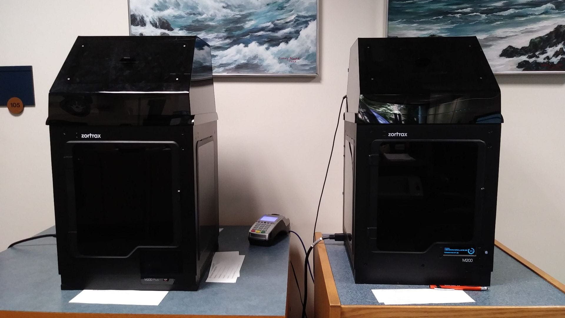
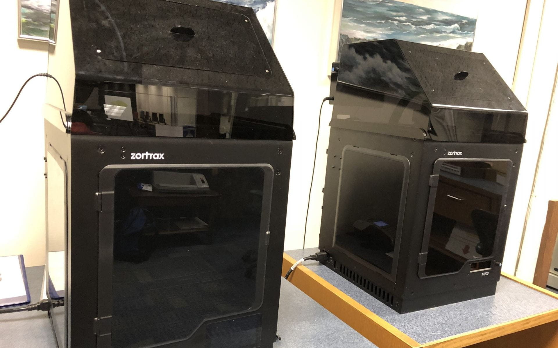
3D Printers on Himmelfarb Library First Floor ©Himmelfarb Library, 2022

3D printing – what is it? It is a method of production, also known as additive manufacturing, in which software guides a machine to craft a 3D object, often with a high level of detail. This, however, is a very generalized definition. It is easy to assume that there is only one kind of 3D printing, but on the contrary: there are many different types of 3D printing, which can be referred to as “processes.” This post seeks to elaborate upon the different 3D printing processes, from the traditional Fused Deposition Modeling that you can find here at Himmelfarb Health Sciences Library, to the more unusual powder bed 3D printing, which is often used in the production of metal objects.

If you difficultly visualizing any of these types of 3D printing processes while reading this piece, check out Horne & Hausman’s 3D Printing for Dummies Chapter 2: Exploring the Types of 3D Printing. This post will describe these processes using more current terms, but this chapter has clear diagrams and illustrations, as well as some additional information about how various 3D printing processes function.

The most popular process of 3D printing is Fused Deposition Modeling, often referred to as FDM printing. This process produces 3D objects by heating up long strings of plastic material (aka: spools of filament), fusing it to the base platform of the printer, then continuing to build it up layer by layer using that same fusion process. Another way of looking at FDM printing is that it’s akin to a highly-nuanced hot glue gun (Horne & Hausman, 2018). There are also variants of FDM printers that utilize pellets instead of filament spools (Volpato et al., 2015), as well as those that use a cold semi-liquid mixture like what you might see with large-scale 3D printed houses that use concrete extrusion (Borg Costanzi et al., 2018).

FDM printing has some limitations. When the filament is in a moldable state during FDM printing, it extrudes through a nozzle, which is almost always moved along X and Y axes like what you might see on a crane game. Because of those movement restrictions, this process is weaker compared to its alternatives when it comes to items that have overhangs or details on an object’s underbelly. The machine is not so sensitive to be able to tell the difference between the 3D printers’ base platform, the previously-placed layers, and open air, so the printer needs the guiding software to give the digital object file (which you can think of as a map or a layout) structures known as “supports” which are designed to be removable. When those supports are removed, it is not uncommon to see score marks on the places where it touched, and in some cases, the area is so narrow that it’s impossible to remove those supports (Horne & Hausman, 2018).

Supports are less of a concern with Stereolithography Apparatus 3D Printing, also known as SLA printing. This 3D printing process produces 3D objects by aiming a laser up into a vat of liquid photopolymerizing resin. Photopolymerizing means that the liquid will transform into a solid when light of a certain wavelength touches it. Unlike FDM printers, which move the entire nozzle horizontally, SLA printers keep the laser fixed in one place, but change the angle. As the software instructs the laser to solidify certain portions of the vat of resin, the object is raised up and out of the vat by a component called the “elevator.” In the case of SLA printers, supports are not needed in order to hold up layers that have overhangs, but instead to keep the object attached to the elevator. SLA printers allow for more complex structures with holes, divots, and overhangs without using up as much filament and without causing scoring marks, and they often manage to print simple objects faster than FDM printers (Horne & Hausman, 2018).

SLA printing has its own downsides to keep in mind. For one, SLA printers tend to be more expensive than FDMs, and they require extra equipment, such as curing stations. Once the object is lifted up and out of the vat, it exists in a semi-cured state and is sticky. It needs to be treated with UV light to finish it. This makes not only cost, but space a concern, particularly since there is liquid resin involved, which requires special storage and disposal methods (Horne & Hausman, 2018).

Powder bed printing, also known as binder jet printing, likewise has some advantages at the cost of specialized set-up requirements. During this 3D printing process, a printer head moves horizontally along X and Y axes like with FDM printing, dropping a liquid binding material onto a powder that covers the printers’ base platform. When the binder comes in contact with the powder, a chemical reaction occurs that solidifies it. Since this process requires the use of fine particles which could be made of metal, plastic, sand, or even plaster, powder bed printers often require hoods and filters to prevent users from breathing in potentially harmful materials. An alternative method of powder bed printing uses a laser in the place of the binding liquid, which burns the powder to solidify it, which adds to the amount of safety equipment required (Horne & Hausman, 2018).

The finished product requires a step known as “depowdering” which can be done by hand with brushes and through automated vibration. Manual depowdering with brushes takes a significant amount of time, whereas automated vibration tools tend to be rather expensive. If you want to learn more about depowdering, this webpage by German depowdering manufacturer Solukon could be of help (Solukon, n.d.).

Powder bed printing is incredibly fast compared to FDM and SLA printing, and it has the added benefit of being able to produce metal objects swiftly. Additionally, this method does not require the use of supports, since the underlying layers of powder that were not activated by the binding chemical or laser can still support the weight of the rest of the object, despite the fact that the head of this kind of printer generally stays on X and Y axes like FDM printers (Horne & Hausman, 2018).

We would be remiss to not mention bioprinting, a process that technically falls under the umbrella of 3D printing which produces natural tissue, often for the sake of testing medications. The process of bioprinting starts by packaging certain types of cells taken from a biopsy into pellets the size of a micrometer or within a liquid that keeps the cells alive. Next, these packaged cells are combined with nutrients and a support material known as a matrix; this combination is known as a bioink. This bioink is then put into place layer by layer by the printer which is guided by software that interprets CT scans and MRIs. Up to this point, bioprinting fits the description of 3D printing, though the materials used are beyond the norm (Wei et al., 2020).

This is where bioprinting diverges, though. Rather than cooling down or solidifying and going off to the post-processing clean-up stage, the cells consume the nutrients and grow within the matrix. Pressure and chemical stimuli are added very carefully to nudge the cells to grow in the intended ways, and chemicals known as bioreactors are added as well to increase the speed of cell growth. There is no kind of 3D printing that is like this, simply because this part of the bioprinting process is less reliant on 3D printing methods and more on natural process of life: consumption, reproduction, and maturation (Wei et al., 2020).

Additional niche 3D printing processes also exist. Laminated Object Manufacturing (LOM) is a process that builds up laser-cut layers of paper, metal foil, or plastic film that has been coated with chemicals of choice. This process uses cheap, readily available materials and allows for additional customization, as the material color can change between layers (Horne & Hausman, 2018). Unfortunately, non-industrial LOM printer manufacturers are few and far between, as they are outpaced in popularity by FDM and SLA printers.

RoboCasting, a method of 3D printing similar to FDM printing which uses a paste made up of materials such as glass or ceramic, which either hardens on its own or needs to be baked. In their work, “3D Printing Bioinspired Ceramic Composites”, Feilden et al. explain how RoboCasting functions and how it can be used to mimic natural materials such as bone and shell (Feilden et al., 2017). This has some benefits for medical sciences through the development of implants such as biodegradable bone scaffolds that can aid during the healing process of bones, which you can learn more about in this study by Lei et al. (Lei et al., 2020).

Each of these 3D printing processes has strengths and weaknesses. Some may be faster, others may be more precise, and others still may be cheaper. This variability makes some more suitable for certain applications than others. FDM, for instance, because of its popularity, limited cost, and low barrier to entry, makes it a perfect choice for early-stage prototyping, whereas powder-bed printing may be more suited for an industrial environment. With 3D printing, the sky is truly the limit, particularly since new printers are being developed each year.

Want to try out 3D printing yourself? We are proud to announce that we have moved to a free-to-print policy and will no longer be charging cost-recovery fees for most print jobs. Some limitations apply, so make sure to consult our 3D Printing at Himmelfarb guide or full policy for the full details.

To learn more about our 3D printing service or to place a request, please visit our 3D Printing at Himmelfarb guide, or contact Brian McDonald (bmcdonald@gwu.edu) for more information.

References:

Borg Costanzi, C., Ahmed, Z. Y., Schipper, H. R., Bos, F., Knaack, U., & Wolfs, R. J. H. (2018)   3D printing concrete on temporary surfaces: The design and fabrication of a concrete shell structure, Automation in construction, 94, p. 395-404 https://doi.org/10.1016/j.autcon.2018.06.013

Feilden, E., Ferraro, C., Zhang, Q., García-Tuñón, E., D'Elia, E., Giuliani, F., Vandeperre, L., & Saiz, E. (2017).  3D printing bioinspired ceramic composites, Scientific Reports, 7(1), p. 1-9. https://doi.org/10.1038/s41598-017-14236-9

Horne, R. & Hausman, K. K. (2018). Exploring the types of 3D printing. 3D Printing for Dummies. https://ebookcentral.proquest.com/lib/gwu/detail.action?docID=4856326 

Lei, L., Wei, Y., Wang, Z., Han, J., Sun, J., Chen, Y., Yang, X., Wu, Y., Chen, L., & Gou, Z. (2020).   Core–shell bioactive ceramic robocasting: Tuning component distribution beneficial for highly efficient alveolar bone regeneration and repair, ACS biomaterials science & engineering, 6(4), p. 2376-2387. https://doi.org/10.1021/acsbiomaterials.0c00152

Solukon. (n.d.). Automating depowdering in 3D printing. Retrieved May 8. 2022 https://www.solukon.de/en/news/festo-and-solukon/

Volpato, N., Kretschek, D., Foggiatto, J. A., & Gomez da Silva Cruz, C. M. (2015)  Experimental analysis of an extrusion system for additive manufacturing based on polymer pellets,  International journal of advanced manufacturing technology, 81(9-12), p. 1519-1531. https://doi.org/10.1007/s00170-015-7300-2

Wei, S., Starly, B., Daly, A. C., Burdick, J. A., Groll, J., Skeldon, G., Shu, W., Sakai, Y., Shinohara, M., Nishikawa, M., Jang, J., Cho, D., Nie, M., Takeuchi, S., Ostrovidov, S., Khademhosseini, A., Kamm, R. D., Mironov, V., Moroni, L., Ozbolat, I. T. (2020). The bioprinting roadmap. Biofabrication, 12(2). https://doi.org/10.1088/1758-5090/ab5158

3D Printers on Himmelfarb Library First Floor
©Leland Ashford Lanquist, 2022

3D printing has received a considerable amount of spotlight in the past decade, but much of the focus lies upon its value for engineering, prototyping, and manufacturing. What can 3D printing offer the field of medical sciences, and what innovations have healthcare professionals already developed using 3D printers? Let’s explore some of the ways 3D printing is able to support or advance the field of medical sciences.

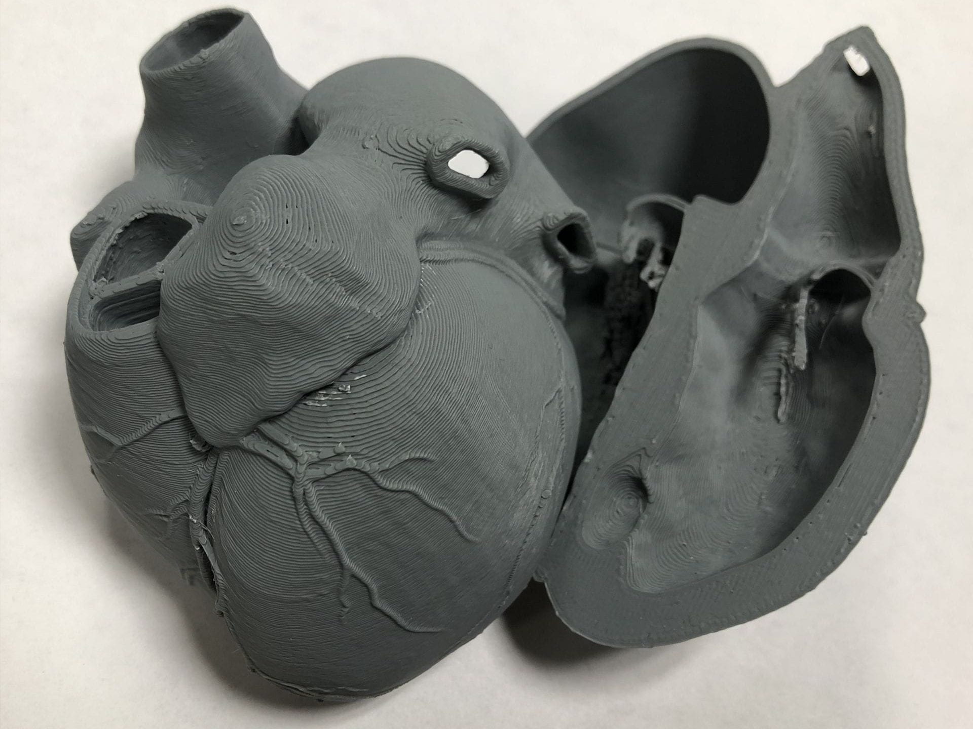
To start with, the customizability of 3D printing offers opportunities to create models that fit the needs of individual students and their courses. Scholars such as AbouHashem et al at Macquarie University and Western Sydney University have studied the effectiveness of using models in education, all at a more affordable price than traditional anatomical models (AbouHashem et al, 2015). But why is 3D printing so much more useful than other approaches to developing educational models? 3D printed models can be developed and printed to suit individual education needs–for instance, one student may want to focus on the internal structures of the heart, but another may need practice with the arteries and veins and how they connect to the rest of the vascular system.

Additionally, while traditional medical models often (for the sake of mass-production) show the body at the peak of health, 3D printed models offer opportunities for physically handling case-study examples. With tools like Harvard’s FreeSurfer, which can transform CT and MRI scans in the form of DICOM files into STL files–files readable by 3D modeling software like Blender3D and AutoDesk Maya–it is possible to create a 3D printable object based off of medical imaging after a bit of work cleaning it up.

3D Printed Heart Model
©Leland Ashford Lanquist, 2022

Aside from educational models, hospitals and medical science institutions have been able to make high-detail models with tactile realism that can be used for surgical training and preparation. One method of 3D printing, known as Fused Deposition Modeling (FDM), extrudes a string of plastic material known as filament through a heated nozzle to meld the layers together one at a time. Advanced FDM printers can use multiple different nozzles to print different colors and materials. This has allowed some professionals such as Watanabe et al to develop models with flexibility and texture that better matches the human body (Watanabe et al, 2021). Weidert et al have elaborated upon how 3D printed models of bone fractures can be used to prepare surgeons for pre-planning complex procedures (Weidert et al, 2019).

Another popularized process is known as “bioprinting.” Bioprinting is a method of 3D printing with cells and other biomaterials to imitate natural tissue. It’s easy to imagine how this could be applied to medical sciences if the technology becomes advanced and accessible enough: as a replacement for organ donation, as a supplement for skin grafts, as a hyper-realistic training tool for surgery preparation. Much of this is not yet feasible on a large scale, but some are becoming more of a reality year by year. For instance, some researchers, such as Keriquel et al, have managed to complete in vivo bioprinting (i.e.: directly printing into the body) bone substitutes in mice with certain bone defects (Keriquel et al, 2017).

If you would like to learn more about bioprinting, Kenneth Douglas’ book, Bioprinting: To Make Ourselves Anew, explains how bioprinting came to be, as well as how it all works, in terms accessible to a generalized audience (Douglas, 2021). For  a deep-dive into where bioprinting as a field might be headed in the future, you may be interested in reading Wei et al’s “The Bioprinting Roadmap,” which analyzes the successes, challenges, opportunities, and obstacles of bioprinting as of 2020 (Wei et al, 2020).

Some surgeons utilize 3D printing to develop customized implants and prosthetics for their patients. Since some of the more high-end 3D printers permit users to print objects made of metal alloys that are safe to be within the human body, healthcare professionals, such as Xu et al, have developed alloy-printed cervical spine reconstruction implants for Ewing’s sarcoma, a rare bone cancer found most commonly in adolescents. 

You can learn more about 3D printed implants by reading Krishna et al’s “Muskuloskeletal 3D Printing” from Rybicki and Grant’s 3D Printing in Medicine. This chapter also includes information about how the customizability of 3D printed prosthetic limbs allows for things such as light-weight, low-cost, and functional prosthetic hands for children that can scale with the natural growth of the patient, (Rybicki & Grant, 2017) which were reported on in more detail by Zuniga et al. (Zuniga et al, 2015) You may also find Christensen’s chapter in 3D Printing in Medicine, “3D Printing and Patient-Matched Implants” a worthwhile read. It covers methods such as using 3D printed patient-scanned models as a form to shape metal implants around prior to surgery, as well as the use of implantable biomaterial, tying in methods of bioprinting previously elaborated upon. (Rybicki & Grant, 2017)

3D printing has not just made advances on a large scale, 3D printing is also on the forefront of innovation within micro-devices like lab-on-a-chip, micro-needles, and more. High-end 3D printers allow researchers and healthcare workers to produce complex micrometer-sized objects such as micro needles and lab-on-a-chip devices, customized to their particular needs.

In terms of the applications of these micro-devices, one scenario might be a researcher using a microelectrode array to gather and track high-quality data about how a person’s muscle cells and neurons react to certain electrical stimuli. This can help pharmacologists better understand how human bodies react to certain drugs. This same device can also be used in the development of a movable prosthetic limb that is custom to the person who uses that prosthesis. 

Microneedles, on the other hand, are tools that allow healthcare professionals to deliver injectable materials into the skin in a way that is less painful and less frightening for patients with needle-phobia. They also produce less waste than their traditional needle counterparts. Researchers such as Kundu et al have published on the value of the production of these kinds of micro-devices in low-resource settings, even despite the high cost of the machines needed to produce them (Kundu et al, 2018). Other researchers such as Santana et al have discussed how micro-devices produced by 3D Printers might serve as a possible alternative to in vivo testing on animals in the future (Santana et al, 2020).

All of this is just a small slice of what 3D printing is capable of in the hands of healthcare professionals. As well, with 3D printing technology advancing, the sky is very swiftly becoming the limit of what is possible. From medical models to research devices, there is so much opportunity that comes with 3D printing for the field of health sciences. 

Want to learn more about 3D printing and even get involved?

Himmelfarb Library offers 3D printing services! While we may not be able to produce every one of the items described in this piece, our 3D printing services do support a wide array of patron projects and activities, from educational models to cookie cutters. It is also a great way to get early involvement with what may very well become standard practice in many aspects of healthcare in the future, so please do come check out the service if you are a patron! If you’d like to learn more about how you can get involved, you can read more about how to request a print in a previous blog post, and you’re welcome to reach out to Leland Ashford Lanquist (lalanquist@gwu.edu) Brian McDonald (bmcdonald@gwu.edu) if you have any questions. If you already know what you want to do, go ahead and submit a print request, which you can also find on our 3D printing guide!

References

AbouHashem, Y., Dayal, M., Savanah, S., & Štrkalj, G. (2015) The application of 3D printing in anatomy education, Medical Education Online, 20(1), https://doi.org/10.3402/meo.v20.29847

Autodesk. (n.d.) Maya Software. Autodesk. https://www.autodesk.com/products/maya/overview

Blender Foundation. (n.d.) Home of the Blender project - Free and Open 3D Creation Software. Blender3D. https://www.blender.org/

Christensen. J. (2017). 3D Printing and Patient-Matched Implants. In F. J. Rybicki & G. T Grant (Eds.) 3D printing in medicine: a practical guide for medical professionals, (pp. 85-95). Springer International Publishing.

Douglas, K. (2021). Bioprinting: to make ourselves anew. Oxford University Press.

Harvard University. (n.d.) Freesurfer. https://surfer.nmr.mgh.harvard.edu/

Himmelfarb Health Sciences Library. (n.d.) 3D Printing at Himmelfarb. https://guides.himmelfarb.gwu.edu/3DPrinting/

Keriquel, V., Oliveira, H., Rémy, M., Ziane, S., Delmond, S., Rousseau, B., Rey, S., Catros, S., Amédée, V., Guillemot, F., & Fricain, J. (2017).  In situ printing of mesenchymal stromal cells, by laser-assisted bioprinting, for in vivo bone regeneration applications. Scientific Reports, 7. https://doi.org/10.1038/s41598-017-01914-x

Krishna, S., Small, K., Maetani, T., Chepelev, L., Schwarz, B. A., & Sheikh, A. (2017). Musculoskeletal 3D Printing. In F. J. Rybicki & G. T Grant (Eds.) 3D printing in medicine: a practical guide for medical professionals, (pp. 71–84). Springer International Publishing.

Kundu, A., Ausaf, T., & Rajaraman, S. (2018) 3D Printing, Ink Casting and Micromachined Lamination (3D PICLμM): A Makerspace Approach to the Fabrication of Biological Microdevices, Micromachines, 9(2), https://doi.org/10.3390/mi9020085

Santana, H. S., Palma, M. S. A., Lopes, M. G. M.., Souza, J., Lima, G. A. S., Taranto, O. P., & Silva, J. L. (2020).  Microfluidic Devices and 3D Printing for Synthesis and Screening of Drugs and Tissue Engineering.  Industrial & engineering chemistry research, 59(9), 3794-3810. https://doi.org/10.1021/acs.iecr.9b03787

Watanabe, N., Yamamoto, Y., Fujimura, S., Kojima, A., Nakamura, A., Watanabe, K., Ishi, T., & Murayama, Y. (2021). Utility of multi-material three-dimensional print model in preoperative simulation for glioma surgery. Journal of Clinical Neuroscience, 93, 200–205. https://doi.org/10.1016/j.jocn.2021.09.017

Wei, S., Starly, B., Daly, A. C., Burdick, J. A., Groll, J., Skeldon, G., Shu, W., Sakai, Y., Shinohara, M., Nishikawa, M., Jang, J., Cho, D., Nie, M., Takeuchi, S., Ostrovidov, S., Khademhosseini, A., Kamm, R. D., Mironov, V., Moroni, L., Ozbolat, I. T. (2020). The bioprinting roadmap. Biofabrication, 12(2). https://doi.org/10.1088/1758-5090/ab5158

Weidert, S., Andress, S., Suero, E., Becker, C., Hartel, M., Behle, M., & Willy, C. (2019) 3D-Druck in der unfallchirurgischen Fort- und Weiterbildung: Möglichkeiten und Anwendungsbereiche, Der Unfallchirurg, 122(6), 444-451. https://doi.org/10.1007/s00113-019-0650-8

Xu, N., Wei, F., Liu, X., Jiang, L., Cai, H., Li, Z., Yu, M., Wu, F., & Liu, Z. (2016) Reconstruction of the Upper Cervical Spine Using a Personalized 3D-Printed Vertebral Body in an Adolescent With Ewing Sarcoma, Spine, 41(1), E50-E54. https://doi.org/10.1097/BRS.0000000000001179

Zuniga, J., Katsavelis, D., Peck, J., Stollberg, J., Petrykowski, M., Carson, A., & Fernandez, C. (2015) Cyborg beast: a low-cost 3d-printed prosthetic hand for children with upper-limb differences, BMC Research Notes, 8(1), 10-10. https://doi.org/110.1186/s13104-015-0971-9