Science & Engineering Hall 5490
❄️The Rodriguez Lab is ready for snow & the holidays!❄️
🎄Decorations include our fluorescent Chemistree, vendor swag tree, poinsettia, candy canes, lights, & holiday cheer!🎄
✨Have a Great Holiday & a Happy New Year!✨#DeckTheLab @Addgene pic.twitter.com/CVD9ZmNnbG— Erik A. Rodriguez (@erin_rod_phd) December 5, 2021
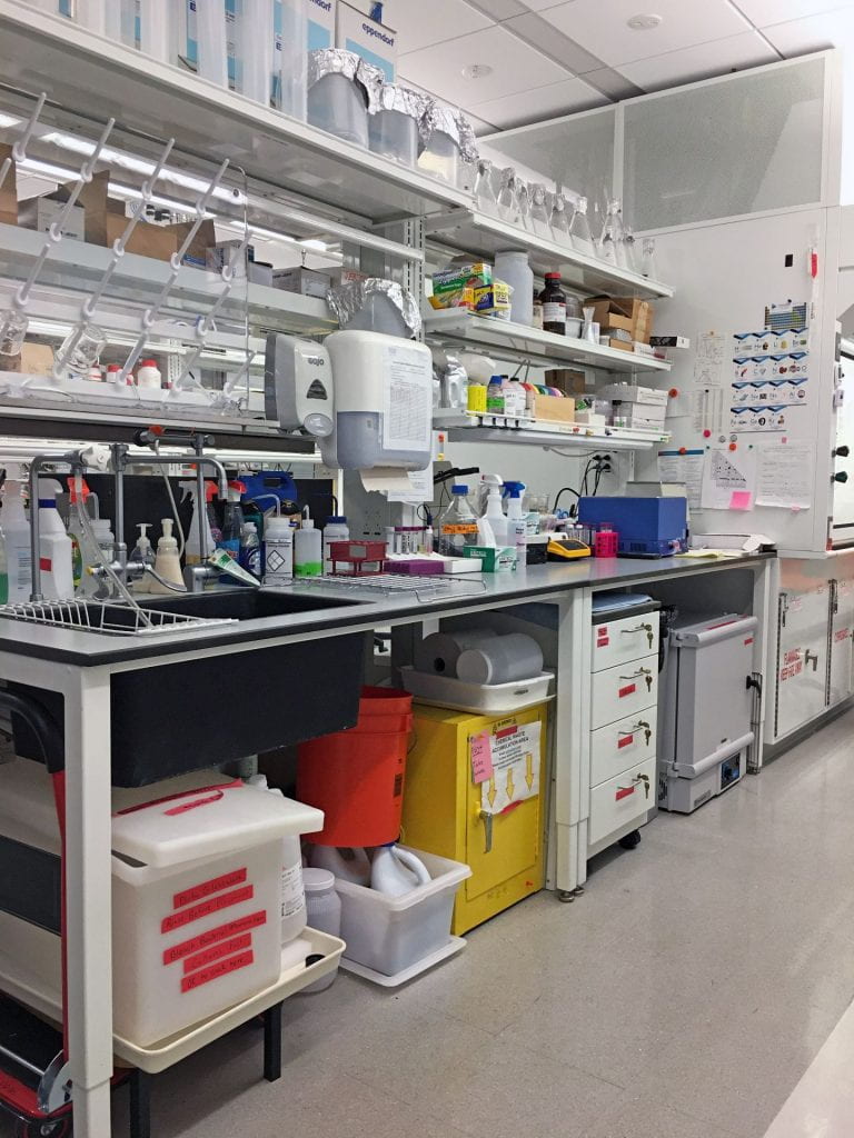
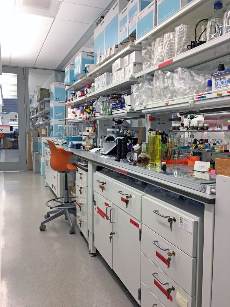
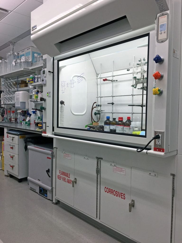
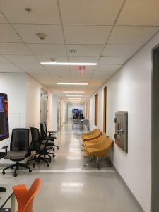
Entrance to the Lab

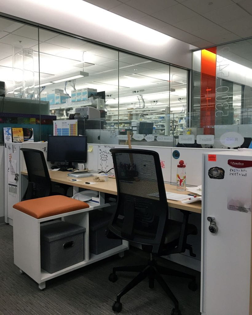
Student Desks

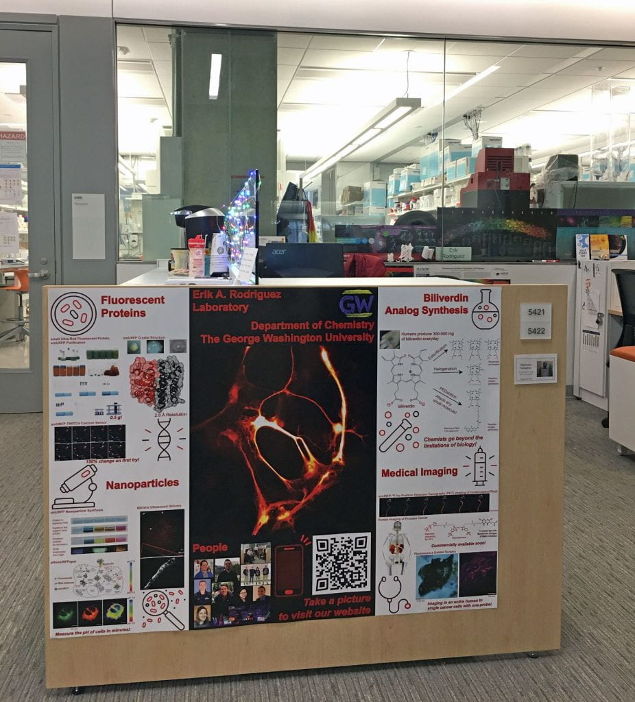
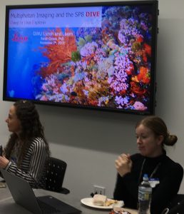
Poster was a winner of the #PostYourPosterChallenge
from The Scientist Magazine (12-2019)!


The view from the #GWU SEH lab is amazing tonight! #4thofJuly2020 #4thofJuly pic.twitter.com/5OIXWndCiy
— Erik A. Rodriguez (@erin_rod_phd) July 5, 2020
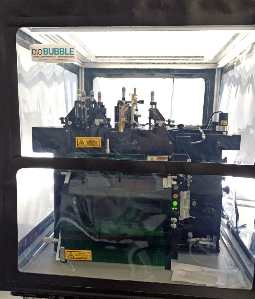
GW Cancer Center Flow Cytometry Core
Fluorescence Activated Cell Sorter (FACS)
BD Influx High Speed Sorter, 4-Lasers, 15-Colors

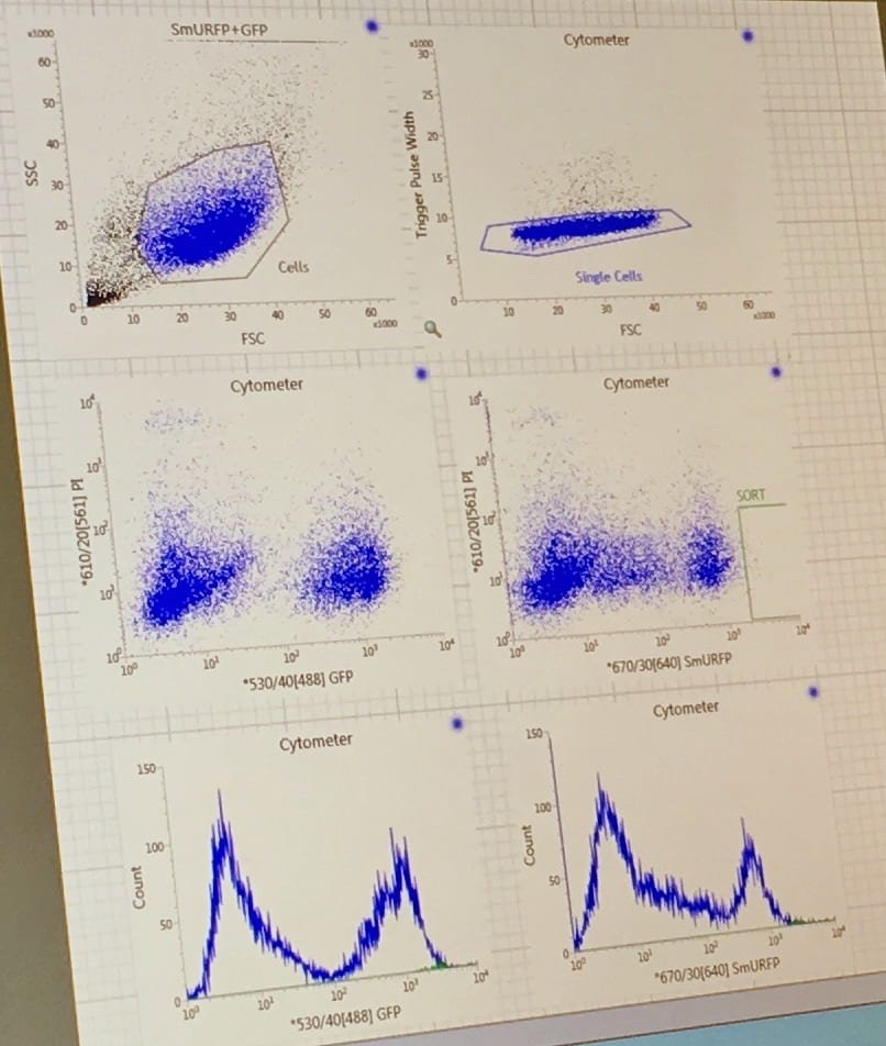
Single cell sorting into a 96 well plate. Profile of HEK293A cells stably expressing
small Ultra-Red Fluorescent Protein (smURFP) and GFP.
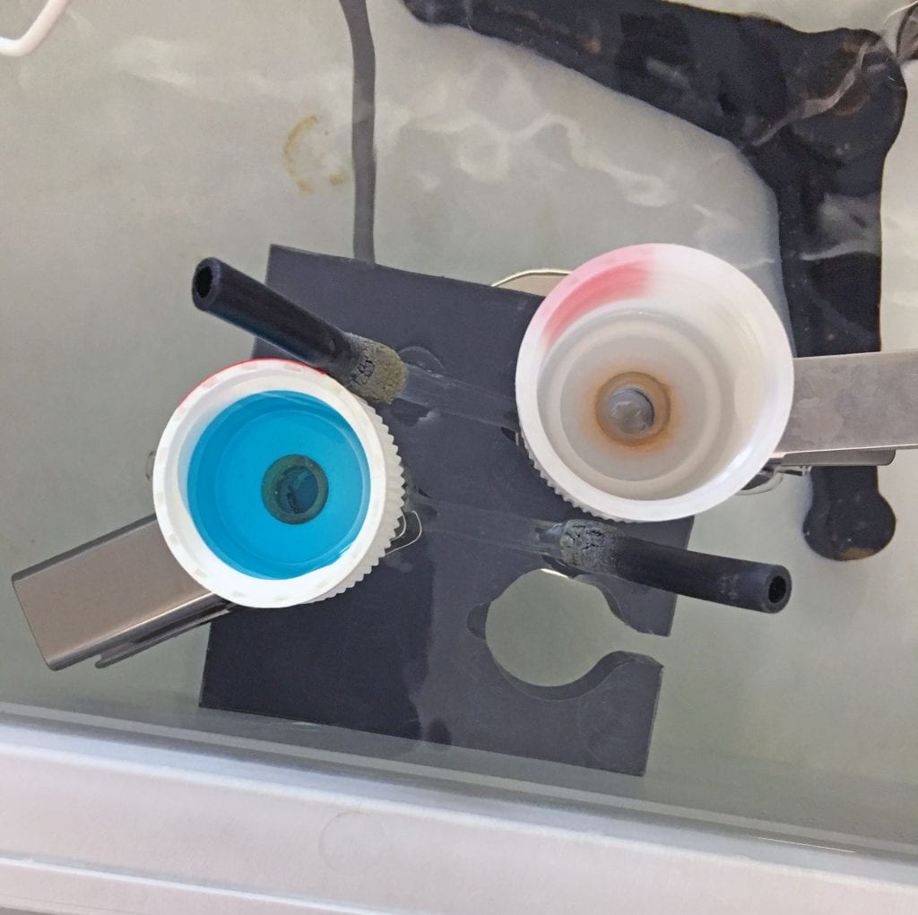
 Ultrasound delivery of small Ultra-Red Fluorescent Protein (smURFP)
& smURFP Nanoparticles.

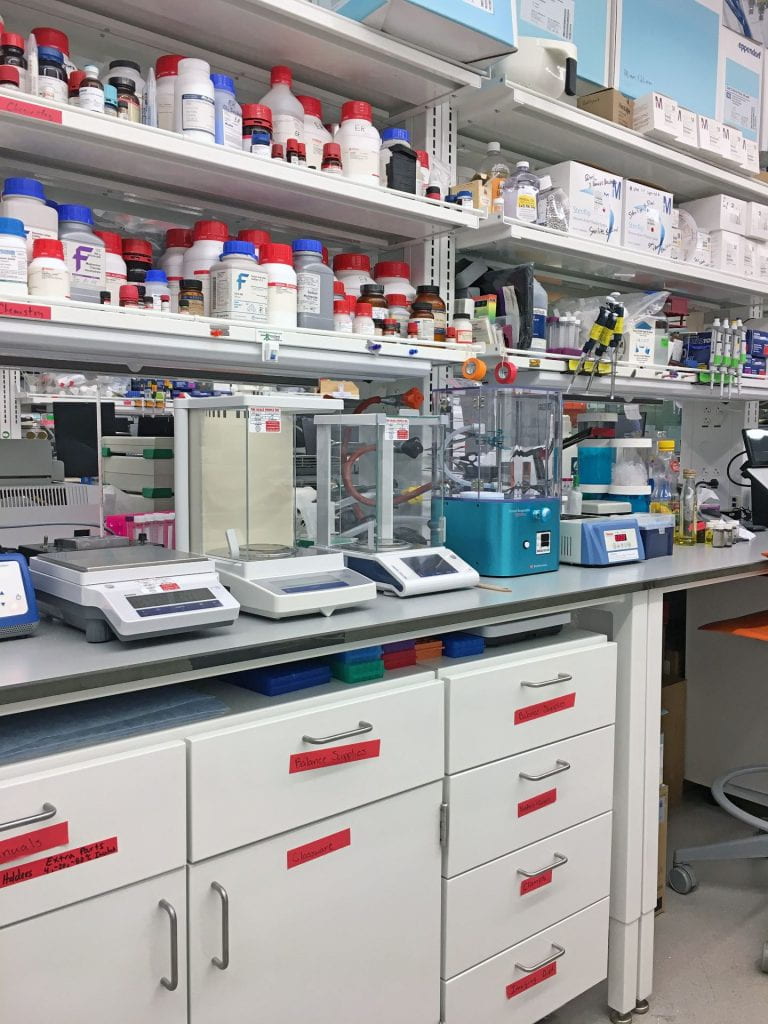
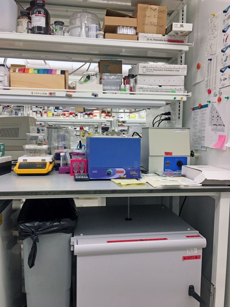
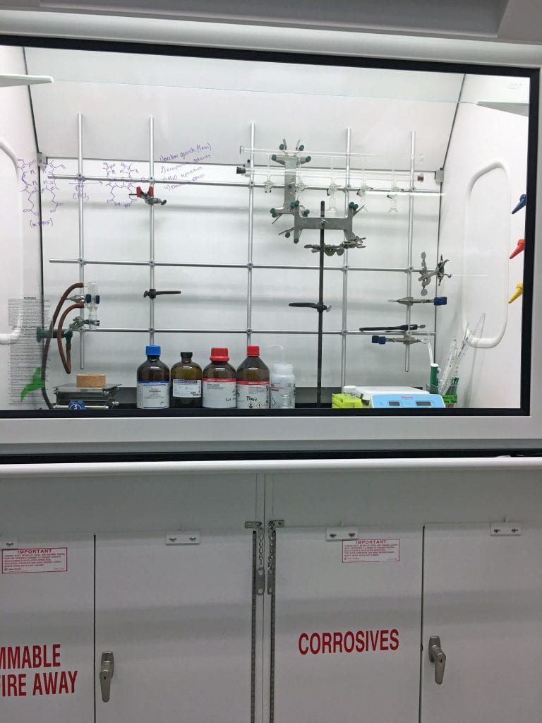
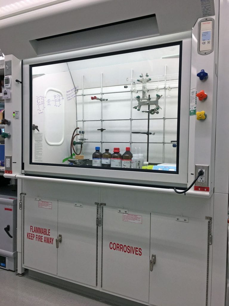
Chemistry Instrument Suite

Instrument Suite: Fluorometer, UV/Vis, FTIR, Raman Spectrometer, ICP-MS with autosampler, GC-MS with autosampler, HPLC with autosampler, Polarimeter, Atomic Absorption Spectrometer, and NMR.
Available to Chemistry Undergraduate, Graduate, and Postdoctoral Researchers when not used by CHEM4123 or other classes.
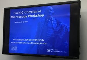
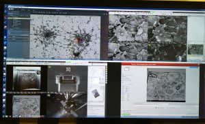
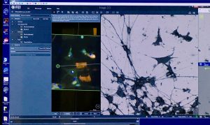
The George Washington Nanofabrication and Imaging Center
Images taken at GWNIC Correlative Microscopy Workshop, Fall 2017.

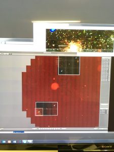
The Google Maps of fluorescence microscopy! 1.2 cm diameter well down to a single cell! Leica TCS SP8 Multiphoton Confocal Microscope-FLIM.
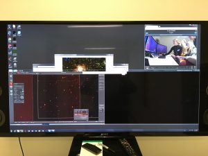
The Leica TCS SP8 Multiphoton Confocal Microscope-FLIM can be controlled remotely!
LeicaMicro EM UC7 Ultramicrotome. Cutting mouse brain slices in 70 nm increments. Macro view of diamond knife trimming the resin.
LeicaMicro EM UC7 Ultramicrotome. Cutting mouse brain slices in 70 nm increments. Eye piece view of diamond knife trimming the resin.
LeicaMicro EM UC7 Ultramicrotome. Cutting mouse brain slices in 70 nm increments. Eye piece view of diamond knife cutting the embedded brain (silver slices in water).
LeicaMicro EM FSP (Freeze Substitution Processor), an automatic reagent handling system. Movie sped up X20 (8 min initial movie to 24 sec in this movie).
LeicaMicro EM Ice. High pressure freezer for CryoEM! Samples are frozen in ≤25 ms in liquid nitrogen! Turn the sound on. The instrument is very loud and pretty shocking.
Electron microscopy images of neurons.

Correlated Light and Electron Microscopy (CLEM)
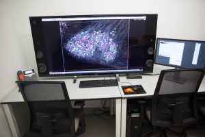
Imaris workstation
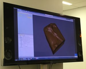
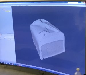 Electron Microscopy of a Milky Way candy bar! Used to determine the air content!