Skip to content

Sonya Chistov

July 6, 2017

A young patient presents to the emergency department (ED) with altered mental status. According to EMS providers, she had ingested an unknown substance, and now was combative, screaming and flailing uncontrollably. Her point-of-care glucose level is normal. She is immediately restrained and sedated, placed in an isolated room and labeled “just another intoxication”. After hours of sleeping, her mental status is not normalizing.  Labs are finally ordered after carefully looking through previous visits and discovering no previous psychiatric history. The patient is found to have an elevated WBC count, is discovered to be febrile and tachycardic. Therefore, what was initially considered a behavioral health issue is actually a medical one that requires immediate care.

Considering the numbers of cases that present similarly to this one, is there a common protocol to distinguish medical from psychiatric emergencies?

Following the closure of institutional psychiatric facilities in the 1980s, EDs are the main location for treatment of patients with a variety of psychiatric complaints [1]. For many behavioral health visits, emergency medicine physicians are commonly tasked with evaluating patients and “medically clearing” them before evaluation by psychiatrists. Medical clearance differs in definition regionally, but often, a set of protocol orders is required before the patient can be further evaluated for evaluation by psychiatrists and potential admission to a psychiatric unit versus a medical unit [2,3]. There has been ongoing debate regarding the necessity of these tests for many patients, because they are often negative and often could potentially be avoided based on clinical grounds.

Each patient in the ED is provided with a physical exam and medical history. If the patient is alert and oriented, expressing only psychiatric complaints, what orders are then deemed necessary for medical clearance?  In many cases, ED physicians will order a basic metabolic panel (BMP), complete blood count (CBC), other functional “psych” lab tests, electrocardiogram (ECG), and a urine drug analysis, as well as an assessment of common overdoses particularly salicylates, acetaminophen, and alcohol. Even with positive urine analysis for drug use, the ED often does not provide further interventions except for time and space to metabolize the ingested agent. [3]

A recent article by Brown et al, in Annals of Emergency Medicine, reviewed numerous studies regarding protocolized psychiatric orders and determined that these tests often had low diagnostic yield, and rarely affected original decisions in disposition. However, the article also noted that psychiatric patients tend to have higher rates of co-morbidities and a shorter general life span, so generic laboratory testing sometimes leads to positive results that may or may not be directly relevant to the ED care. Patients also spend long periods of time in the ED before evaluation, until the decision for admission is made, and until they receive a hospital bed. [4,5,6] Those in favor of the standardized testing work closely within the medical model. These people suggest that underlying illness can influence psychiatric complaints, and these tests can determine or eliminate a direct or organic medical cause. Also, due to overcrowding on inpatient psychiatric units, the use of these tests can weed cases that may be more appropriate for “medicine” cases, saving room for true psychiatric emergencies. [4,5,6]

The lack of consensus regarding all aspects of this issue is astounding, but ultimately it comes down to two things: time and money. If patients complaining of new onset psychiatric complaints, with no other previous medical history, are evaluated the same as patients with a history of psychiatric complaints, substance abuse, or extensive co-morbidities, is it necessary to be “treated” in the ED? In my humble opinion, our healthcare system should develop a standardized protocol, which can compromise between both sides of this argument. If a patient presents with psychiatric symptoms, they should be examined as any other patient with a routine physical exam and obtaining a detailed medical history. If the patient has stable vitals, normal physical exam, no substance abuse (altered mental status) and no other complaints- the complaint could potentially be evaluated directly by psychiatrists or within a psychiatric-focused ED, as many hospitals have this already. For patients deemed a higher risk, such as elderly patients, patients with new onset psychiatric symptoms and patients with altered mental status, testing should be determined based on each patient’s presentation and medical necessity, and by the treating ED physician.

References:

[1] Larkin GL, Claassen CA, Emond JA, Pelletier AJ, Camargo CA. Trends in US emergency department visits for mental health conditions, 1992 to 2001. Psychiatric services. 2005 Jun;56(6):671-7.

[2] Brown, MD. et al. Clinical Policy: Critical Issues in the Diagnosis and Management of the Adult Psychiatric Patient in the Emergency Department. Annals of Emergency Medicine. 2017, Feb; 69(4): 480-98

[3] Turner JE, Zun LS. An Evidence-Based Approach to Medical Clearance of Psychiatric Patients in the Emergency Department. Current Emergency and Hospital Medicine Reports. 2015 Dec 1;3(4):176-82.

[4] Tucci V, Siever K, Matorin A, Moukaddam N. Down the rabbit hole: emergency department medical clearance of patients with psychiatric or behavioral emergencies. Emergency medicine clinics of North America. 2015 Nov 30;33(4):721-37.

[5] Anderson EL, Nordstrom K, Wilson MP, et al. American Association for Emergency Psychiatry Task Force on Medical Clearance of Adults Part I: Introduction, Review and Evidence-Based Guidelines. Western Journal of Emergency Medicine. 2017;18(2):235-242.

[6] Zun L. Care of psychiatric patients: The challenge to emergency physicians. Western journal of emergency medicine. 2016 Mar;17(2):173.


Sonya Chistov is an ED Technician at The George Washington University Hospital

1

Rose Kleiman

June 26, 2017

Is the emergency department (ED) the right place to inquire about a patient’s sexual orientation? A recent study published in JAMA Internal Medicine points toward yes[i].

While lesbian, gay, bisexual, transgender, and queer (LGBTQ) populations often report poorer health and less access to health insurance and health services, lack of data on sexual orientation is a barrier to understanding and addressing these disparities. Recent efforts to capture data on sexual orientation have been made by both the US Department of Health and Human Services and the National Academy of Medicine. Yet, few health systems or emergency departments (EDs) regularly collect data on sexual orientation.

Researchers for the Emergency Department Query for Patient-Centered Approaches to Sexual Orientation and Gender Identity (EQUALITY) Study used a mixed-methods approach to try and understand the willingness of patients to disclose and of providers to collect information on sexual orientation in the ED. The research team conducted in-depth interviews with patients and ED professionals in the Baltimore, Maryland, and Washington, DC, areas and used results from the first, qualitative phase to develop a national survey of patients and ED providers.

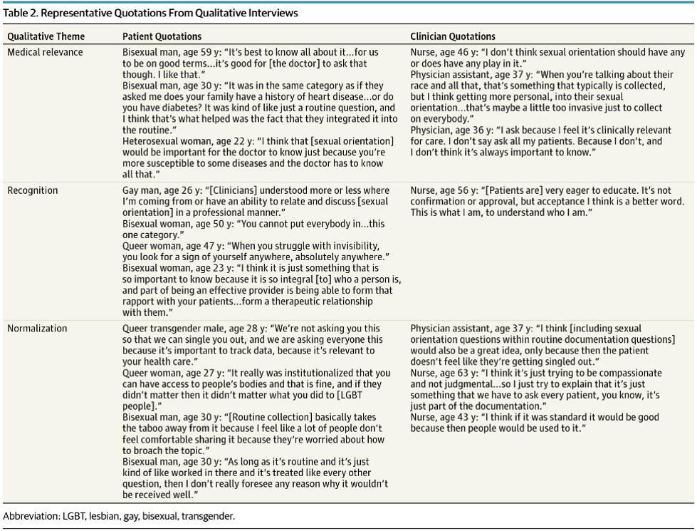
Findings from their qualitative interviews (see Table 2 below) highlight the following themes when discussing routine collection of sexual orientation in the ED: Medical relevance, normalization, and recognition.

The qualitative interviews describe the disconnect between patients and providers. Emergency providers felt if sexual orientation was not relevant for a patient’s immediate care plan, it was not necessary to know and thus not important to ask. Patients felt that their sexual orientation was essential information for their overall health and wellness, similar to inquiring about family history of heart disease or exercise habits, and thus should be a routine part of screening. Therefore, the lack of screening on sexual orientation may be a missed-opportunity for providers to build meaningful relationships with patients.

Similar findings were reflected in the quantitative results from the national survey. A total of 80% of emergency providers reported they thought patients would be offended if asked their sexual orientation in the ED, with 78% believing patients would refuse to provide this information. By contrast, only 10% of patients reported they would refuse to provide such information in the ED and only 11% reported that they would be offended if sexual orientation data were routinely collected.

Patients in the survey also emphasized the importance of collecting data on sexual orientation for recognition and normalization of LGBTQ individuals in society. It was expressed that standardizing the collection of this information may help to further promote patient-centered care for all patients.

The research team found that the preferred method of both patients and clinicians for collecting this information was through a nonverbal self-report. The EQUALITY team currently has a trial under way to study the different ways of collecting this information to determine the optimal method.

This study does have some limitations. First, the qualitative interviews were only sampled from one region of the United States; however, the interviews informed the development of the survey, which found similar themes on a national level. In addition, the study did not test how patients actually respond when asked about sexual orientation information in a clinical setting.

While this article highlights the significance of collecting data on sexual orientation, it leads to an important message for emergency providers: patients believe that their sexual orientation is an important component of their overall health and feel that it is necessary information for their providers to know. Whether it is essential to determining an immediate treatment plan or not, querying about a person’s sexual orientation can lead to a more person-centered approach to care.

[i] Haider, Adil H., et al. "Emergency Department Query for Patient-Centered Approaches to Sexual Orientation and Gender Identity: The EQUALITY Study." JAMA Internal Medicine (2017).


Rose Kleiman is a medical student at the GW School of Medicine & Health Sciences

Evan Kuhl, MD

June 15, 2017

Radiographic contrast media is considered a common cause of hospital-acquired renal insufficiency, yet the latest research on contrast-induced nephropathy (CIN) suggests there may be no relationship between contrast use and renal injury1,2. A recent article by Dr. Hinson et al published in the Annals of Emergency Medicine, found no association between acute kidney injury and IV contrast use. Hinson’s article, a single-center retrospective cohort analysis of 17,934 patients who underwent CT scan with or without contrast, is the largest controlled study of CIN, and may lead to a significant change in practice.

Contrast-induced Nephropathy is a change in creatinine of 25% from baseline, or absolute increase of 0.5 mg/dl 48-72 hours after infusion1. Early research identified CIN in patients undergoing coronary catheterization, where much larger loads of contrast was used, and before the use of low-osmolar contrast agents3. Other research has found that underlying renal disease, or already impaired renal function, may be the cause of CIN4. CIN may be due to many factors other than IV contrast. Patients undergoing a CT with IV contrast followed by a rise in creatinine may have alternative causes which are overlooked, such as nephrotoxic drug use, sepsis, or a range of other causes. The contrast is presumed to be the cause, and much of the research surrounding CIN is hindered by confounding bias due to the exposure itself and the physiology of why the patient is receiving the scan in the first place. Measuring the creatinine level in patients undergoing multiple procedures, nephrotoxic medication use, or requiring CT imaging creates a significant selection bias. When selection bias is accounted for with negative controls, renal injury rates have not been correlated with contrast use, or associated with previous renal injury5,6.

The most recent study by Hinson sought to clarify the incidence of CIN and utilized two control populations to minimize bias. The paper was a retrospective review of 17,934 patients over a 5-year period, comparing the incidence of AKI in contrast-enhanced CT, non-contrast CT, and no CT imaging, with the primary outcome of incidence of acute kidney injury. Patients were included if they were >18 years-old, received an CT scan either with or without contrast, and had both initial creatinine and 48-72 hour creatinine measurements. A second control group was also included who did not undergo CT imaging, but did have initial and secondary creatinine measurements. Patients were excluded if they already had severe renal injury, a prior renal transplant, recent prior CT imaging, or insufficient creatinine data. The study used both the classic definition of CIN, as well as the Acute Kidney Injury Network/Kidney Disease Improving Global Outcomes (AKIN/KDIGO) guideline to define renal injury as a primary outcome.

The results found that contrast used produced no significant increase in acute renal injury. Using AKIN/KDIGO scoring, the probability of AKI was 6.8% in those with contrast-enhanced CTs, 8.9% in non-contrast CTs, and 8.1% for those without any CT imaging. Classic CIN definitions found the probability of developing AKI in each group was 10.6%, 10.2%, and 10.9%, in contrast-enhanced, non-contrast, and no CT imaging groups respectively. There was also no significant change in the risk for developing chronic kidney disease in 6 months or initiating dialysis between the groups.

Although this study is retrospective and single-center, it represents the largest controlled study of CIN. The inclusion of two control groups reduces the selection bias, and the primary outcome was reported using both AKIN/KDIGO and classic CIN definitions, preventing any under-reporting of renal injury. While the article found no evidence of contrast-induced nephropathy, they do identify some nephroprotective treatment patterns as being potentially responsible. Providers were more likely to provide intravenous fluids and less likely to order contrast-enhanced studies to those with decreased renal function or comorbidities. With these results, the authors are calling for a controlled randomized prospective trial to determine if there is a causative relationship between contrast media and acute renal injury.

1.         Hinson JS, Ehmann MR, Fine DM, et al. Risk of Acute Kidney Injury After Intravenous Contrast Media Administration. Ann Emerg Med 2017;69:577-86 e4.

2.         Nash K, Hafeez A, Hou S. Hospital-acquired renal insufficiency. Am J Kidney Dis 2002;39:930-6.

3.         Daniel J. Pallin M, MPH. Intravenous Contrast May Pose No Risk to Kidneys. 2017.

4.         Pickering JW, Blunt IR, Than MP. Acute Kidney Injury and mortality prognosis in Acute Coronary Syndrome patients: A meta-analysis. Nephrology (Carlton) 2016.

5.         Contrast-Induced Nephropathy: Confounding Causation - emDOCs.net - Emergency Medicine Education. 2017. at http://www.emdocs.net/contrast-induced-nephropathy-confounding-causation/.)

6.         Davenport MS, Khalatbari S, Cohan RH, Dillman JR, Myles JD, Ellis JH. Contrast Material–induced Nephrotoxicity and Intravenous Low-Osmolality Iodinated Contrast Material: Risk Stratification by Using Estimated Glomerular Filtration Rate. Radiology 2013;268:719-28.


Evan Kuhl, MD is an Emergency Medicine Resident at The George Washington University Hospital

1

Austin Wu

June 7, 2017

As described in a previous Urgent Matters blog post, opioid prescriptions in the emergency department (ED) have the potential to cause long-term opioid use (defined as 180 days or more of opioids within 12 months of the index ED visit). Further, prescription opioids continue to be the number one cause of drug overdose deaths in the US.[1] These trends indicate a dire need for effective interventions to curb unnecessary opioid prescriptions and prevent opioid abuse by patients.

A recent study in the Annals of Emergency Medicine examines the effects of one such intervention: opioid prescribing guidelines. Specifically, the study examined opioid prescription rates by ED physicians in Ohio, comparing pre and post guideline data.[2]  The goal was to determine whether the implementation of Ohio’s April 2012 opioid prescription guidelines for ED physicians reduced the number of opioid prescriptions by ED physicians.

Researchers utilized data from Ohio’s prescription drug monitoring program from 2010 – 2014, and conducted an interrupted time series analysis. Ohio’s Prescription Drug Monitoring Program includes all prescriptions for schedule II to IV medications dispensed by a pharmacy within the state. Prescriptions written in Ohio but dispensed outside the state were not included. The 5 most commonly prescribed opioids (hydrocodone, oxycodone, tramadol, codeine, and hydromorphone) were examined in this study, and prescriptions counted were limited to those prescribed by physicians with a primary specialty of emergency medicine, pediatric emergency medicine, and sports medicine. Orthopedic surgery opioid prescriptions were utilized as a control against confounding variables, primarily from opioid-related interventions initiated in parallel with the new guidelines.

Three measurements were followed in the study: 1) Total opioid prescriptions in Ohio per month by ED physicians 2) Total morphine milligram equivalents contained in these monthly prescription totals 3) Number of opioid prescriptions greater than 3 days’ duration (specifically discouraged by guidelines). The data in January of 2010 showed that total opioid prescriptions dispensed by all ED physicians in Ohio declined by 0.31% per month, which than changed to a 11.98% decrease per month after guideline implementation, starting April of 2012. Total morphine milligram equivalents improved from a 0.29% decrease per month to 17.36% decrease per month. Opioid prescriptions greater than 3 days’ duration improved from 0.04% per month to 11.2% per month. Further, the rate of decline in all three of these measures continued to decline at approximately 0.9% per month. The graphs below depict total prescription trends when stratified into the 5 most commonly prescribed opioids. A, B, C, D, and E represent Hydrocodone, Oxycodone, Tramadol, Codeine, and Hydromorphone, respectively.


SOURCE: Scott G. Weiner et al., “The Effect of Opioid Prescribing Guidelines on Prescriptions by Emergency Physicians in Ohio,” Annals of Emergency Medicine, May 2017, doi:10.1016/j.annemergmed.2017.03.057.

As demonstrated by the downward shift after the May 2012 time point, all five opioids displayed a reduction in number of prescriptions after the guidelines were implemented. The steeper slopes also demonstrate an increased rate of decline in opioid prescriptions post-guidelines.

There were several limitations to this study, including possible data entry errors, a focus on a specialty representing only 5% of total opioid prescriptions, and limited generalizability as a statewide study. However, this research suggests promise for using guidelines, both within and outside the specialty of emergency medicine, as one effective tool for combating the opioid epidemic.

[1] Rose A. Rudd, “Increases in Drug and Opioid-Involved Overdose Deaths — United States, 2010–2015,” MMWR. Morbidity and Mortality Weekly Report 65 (2016), doi:10.15585/mmwr.mm655051e1.

[2] Scott G. Weiner et al., “The Effect of Opioid Prescribing Guidelines on Prescriptions by Emergency Physicians in Ohio,” Annals of Emergency Medicine, May 2017, doi:10.1016/j.annemergmed.2017.03.057.


Austin Wu is a medical student at the GW School of Medicine & Health Sciences

Will Denq, MD & Evan Kuhl, MD

May 31, 2017

In 2016, an interesting and potentially practice-changing articles was published: the PESIT trial by Prandoni et al.. The prospective study utilized 11 Italian hospitals and evaluated all patients for pulmonary embolism (PE) who were admitted to the hospital for first-time syncope. The results made for headlines; pulmonary emboli were found in 1 in 6 patients, and was broadcast by major news outlets including NPR and Medscape. With the headlines came a debate amongst physicians about just how useful--if not harmful this study might be.

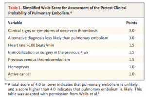
Prior to the PESIT trial, there was scant data on the prevalence of PE in patients admitted for syncope. The authors created a multicenter, cross-sectional study with 11 hospitals in Italy to evaluate the frequency of PEs in patients admitted for syncope. Each patient was risk stratified with a Simplified Wells Score (see Table 1) Those with a score greater than 4 or with a positive D-dimer underwent CT angiography or V/Q scan to evaluate for pulmonary emboli.

Of 2,584 patients who were evaluated by the Emergency Department for syncope, 560 patients were included in the study. Of these, 230 patients received CTA or V/Q scans, and 97 patients were found to have PEs.

The Objective Results:

  1. 2584 patients assessed
    1. 560 included (Mean age = 76 )
    2. 1867 patients discharged and therefore excluded
    3. 157 inpatients further excluded
      1. Prior episodes of syncope, anticoagulated, pregnant excluded
  2. 97/560 (17.3%) were found to have PE
    1. 230/560 were high risk -> CTA or V/Q scan
    2. 180 had CTA, 1 had autopsy
      1. 73/180 had PE
        1. 49/73 (67%) had a main pulmonary artery or lobar artery PE
        2. 19/73 (26%) had segmental PE
        3. 5/73 (7%) had subsegmental PE
    3. 49 had V/Q
  3. 330/560 were low risk -> no imaging

The result of this study carries a bold statement. 1 in 6 of patients hospitalized for syncope have a PE, therefore perhaps we should consider testing syncope patients for PE even when we think the symptoms are caused by something else given the high prevalence of disease.  However, without reading the paper in detail may result in lowering the threshold to test for PE and potentially an overutilization of D-dimer, CTA, and/or V/Q scans. Below,we present the pros and cons of this paper to better inform whether to consider PE in your next syncope patient.

Practice Changing:

Bottom Line: The PESIT trial is a well designed study that should increase our consideration of PE during syncope workups, especially in the geriatric population. When a pulmonary embolism causes syncope, a missed diagnosis may cause significant complications and patient safety issues.

Although we are getting better at catching PEs, they can often be subtle and difficult to diagnose. If a casual reader were to browse through the abstract or to base their judgement on a tertiary news source, they would miss the true value behind this study. It actually calls for a closer examination of PE in older patients that are admitted for first-time syncope, not for testing everyone. Those who are low risk and considered safe for discharge were not included in this study, and this study should not impact our practice in these patients. Those at high enough risk to warrant admission may need a second look. In those with prior PEs, undetermined cause of syncope, or clinical features of PE (respiratory distress, tachycardia, hypotension, or signs of DVT) we should have an increased suspicion of pulmonary embolism.

This conclusion results from the question of, how exactly do we translate an inpatient study and apply it to the ED setting? What population of syncope seen in the ED do we even consider PE in? Based on the population you see and ED you work in, admission criteria for syncope is likely to differ. For appropriate reasons, there wasn’t a standard for syncope admission in this study. However, if we refer to Table 1 of the article, a useful data point is the age of the study population. Not so surprisingly, the mean and median age of the study population was 76 and 80, respectively. It makes sense. Geriatric patients tend to have higher admission rates since they have more comorbidities, less reserve, and a larger number of possible etiologies for their symptoms.

Although there is a different discussion to be had about the causality, it is still standard of care to treat PE even if it exists as a non-etiologic diagnosis for their syncope admission. 67% of patients with a CT positive for PE had a significant embolus that could be related to the syncope they experienced. Only 7% of CT-PE positive patients had subsegmental PEs. The best we can take from this data is to consider PE as an association in the geriatric population that presents as first-time syncope.

Not so fast:

Bottom Line: This inpatient study has the dangerous implication of resource overutilization and the results are difficult to translate to other countries and the ED.

There are radiation and potential contrast risks, workflow, and healthcare costs to consider especially if patients or providers do not understand the implications behind this study. The study does not imply that 1 in 6 patients syncopized because of a PE. It just reports that of the patients admitted for syncope, 1 in 6 were found to have a PE. Association, but not causality is what this is. 12.7% of those found to have pulmonary emboli had already been given an alternative diagnosis.  Many of these patients may have presented to the ED with an emboli, and others may have been asymptomatic as the clinical significance of these findings is not known.

While the authors focus on the number of patients admitted, they do not account for the 2584 patients screened in the ED. Additionally, many of the patients found to have PEs showed clear evidence of DVT and would have a high Well’s score, which should be identified in the ED prior to admission. When all 2584 patients are taken into consideration, you see a much more typical rate of PEs that many of us are more used to seeing, near 5-10%. It is difficult to translate the algorithm and results of the study across the world from Italy to the US. Do we also discharge 72% of our first time syncope patients? Do we D-dimer all of our syncope patients? The clear answer is no. Despite the authors’ claim that the “standardized protocol” for the diagnostic workup of syncope was based on international guidelines - there is no mention in either guideline about the use of D-dimer or even Well’s in the initial consideration for syncope (1,2).

Summary:

Bottom Line: This study is not practice-changing, but it should increase our awareness for the prevalence of PE in the geriatric population that presents with first-time syncope. If you decide to change your practice, consider the downstream consequences.

There may be some benefit in considering PE in the differential in the admitted geriatric patient with first-time syncope. However, the guidelines (1,2) from both cardiology societies in the United States and Europe do not even discuss the use of D-dimer or Well’s criteria in the initial workup for syncope. Although these tools are an acceptable method for ruling out PE, PE may not even be on the differential for a first-time syncope patient.

This study potentially opens a can of worms. If we start to screen the geriatric population more for PEs, what are the downstream consequences? We will begin to adopt age-adjusted D-dimers more? Will we seen an increase in V/Q scans in patients with kidney disease? Will we see an increase in kidney disease from the contrast used in CTAs? What is the appropriate management for the subsegmental PEs that we find? Will we see an increase in bleeding complications such as GI bleeds or head bleeds in the fall-prone geriatric population? What implications will this have for ER workflow? This article, like many others, is merely a beginning step towards more inquiry. It is not practice-changing, but should increase awareness for the prevalence of PE especially in our geriatric population.

What this paper does do is to open an avenue to have discussions with patients. In the world of the internet and immediate access to information, patients come with new expectations. While we may see an increase in patients or other physicians requesting CT scans and D dimer testing, we have the opportunity to better inform our patients. Identifying your patient’s concerns and addressing them with evidence-based medicine should be the foundation of our practice. Being able to translate the evaluation for DVT and PE for a lay person can be difficult, but may produce a more informed and empowered patient. This paper, and the media attention it received, is a launch point for a larger discussion we should all be having with our patients.

  1. Task Force for the Diagnosis and Management of Syncope, European Society of Cardiology, European Heart Rhythm Association, Heart Failure Association, Heart Rhythm Society. Guidelines for the diagnosis and management of syncope (version 2009). Eur Heart J 2009;30:2631-2671
  2. Strickberger SA, Benson DW, Biaggioni I, et al. AHA/ACCF Scientific Statement on the evaluation of syncope: from the American Heart Association Councils on Clinical Cardiology, Cardiovascular Nursing, Cardiovascular Disease in the Young, and Stroke, and the Quality of Care and Outcomes Research Interdisciplinary Working Group; and the American College of Cardiology Foundation: in collaboration with the Heart Rhythm Society: endorsed by the American Autonomic Society. Circulation 2006;113:316-327


Will Denq, MD & Evan Kuhl, MD are both Emergency Medicine Residents at The George Washington University Hospital

Ameer Khalek

March 21, 2017

Patients presenting with chest pain associated with normal EKGs, negative cardiac enzymes, and few cardiac risk factors are designated “low-risk,” and can often be safely to discharge from the emergency department (ED) for early patient follow-up if no emergent conditions are found. ED care for low-risk chest pain is common: United States EDs annually see over 8 million visits for chest pain or related symptoms. Since chest pain is a high-risk complaint (i.e., imminent threat to life), a large amount of resources is allocated to detect and treat cardiopulmonary disease. Missed myocardial ischemia is clinically important concern: failure to detect it leads to a risk-adjusted mortality ratio that nearly doubles that of patients hospitalized for the same condition. An earlier Urgent Matters post by Dr. Evan Kuhl discussed the concept of shared decision making in patients with low-risk chest pain, which describes a way that providers can communicate risk with patients and deliver care in accordance with their values.

After ensuring no serious medical problems are present, the question for ED physicians and patients is: “What is the actual cause for the symptoms and how should they be treated?” Prior studies have shown that up to 55% of patients with non-cardiopulmonary chest pain may be suffering from anxiety or panic disorders, which remain undiagnosed in almost 90% of cases (Foldes-Busque et al & Eken et al). A study by Musey Jr. et al in press in The Journal of Emergency Medicine examines for self-reported stress/anxiety in low-risk chest pain patients. Dr. Musey Jr.’s study is a secondary analysis of prospective outcomes from four centers including 851 participants presenting to the ED with chief complaints of chest pain and or shortness of breath. Participants were divided into two groups (1) explicitly self-reported anxiety/stress (2) no explicitly self-reported anxiety/stress at 90-day follow-up. These groups were compared on several metrics including pretest probability (PTP), outcome rates for ACS and pulmonary embolism (PE), radiation exposure, and 90-day recidivism. When asked “What do you think caused your chest pain?” sixty-seven (8%) patients responded explicitly that their chest pain was caused by mental “stress” or “anxiety.” From this group, the mean ACS PTP was 4% (95% [CI] 2.9-5.7%), with 49% (33/67) having an ultralow (<2.5%) ACS PTP. The mean PE PTP was 5% (95% [CI] 3.6-5.7%) with 46% (31/67) having an ultralow PE PTP. The study found that none of the 67 patients had ACS or PE, and their radiation exposure, costs associated with care, and recidivism rates were similar to patients who did not volunteer anxiety as the cause of their chest pain.

Additionally, none of the patients with self-perceived anxiety were treated for anxiety or received a diagnosis of anxiety.

From the entire cohort, only two patients were given an ICD-9 diagnosis of anxiety. Musey Jr. et al.’s findings may indicate a missed opportunity to detect a treatable condition in patients who seldom receive an actionable diagnosis as opposed to descriptive “chest pain.”

“Detection and treatment of anxiety and panic syndromes may help forestall development of chronic chest pain and recurrent desire for medical reassurance, despite negative cardiac evaluations.”

Ultimately, we need to focus on treating not only the serious but also the non-serious symptoms for ED patients. This is especially true in chest pain given the recent literature on the topic.


Ameer Khalek is a MPH student at the GWU Milken Institute School of Public Health

Mohammed Alabdrab Alnabi, MD

May 18, 2017

Emergency care for sepsis has evolved considerably over the past two decades. Prior to the Rivers trial in 2001, antibiotics were the mainstay of treatment, which then evolved into early resuscitation with intravenous fluids, blood products, invasive monitoring, and vasopressors.(1) In the past year, results from the Process, Promise, & Arise trials, then their recent PRISM meta-analysis challenged the concept of the important of invasive monitoring, but still underscore the benefits of early antibiotics and aggressive resuscitation (2,3,4,5).

Through this process and evolution, there has been a dramatic reduction in sepsis related mortality. Current approaches to sepsis resuscitation focus on the host by reducing hypo-perfusion. However, few effective therapies focus on the host response.Efforts to impact the host response have been disappointing after initial success, specifically with using steroids and activated protein C to treat sepsis (6,7, 8,9,10).

In recent months there has been a new development in sepsis, specifically a recent study by Paul Marik, published in CHEST. (11) Marik’s study tests a "sepsis cocktail", consisting of vitamins C & B1, combined with steroids, along with standard treatments for sepsis. In the trial, each of the components of his cocktail has been tested individually before, and showed some promise. For example, burn patients who receive Vitamin C require less fluids & produced more urine (12). Similarly, surgical ICU patients who were given Vitamin C had less ventilator days & less multiorgan failure (13). Patients with severe sepsis who were given Vitamin C had better SOFA scores (14), reduced mortality, & reduced vasopressin use (15). Thiamine too, has been shown to reduce mortality in septic shock among patients with thiamine deficiency (16). The addition of steroids in Marik's study was probably because of its synergistic effects with vitamin C (17).

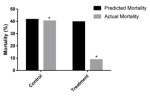
In the Figures, we see quite dramatic results with a great reduction in mortality in treatment as compared to the control arm, albeit this was a before-after study with historical controls.  In addition, we see a significant reduction in SOFA scores – a measure of severity of illness. But before we say "Eureka", we need to carefully consider how to interpret these results. In the past, we have been tricked before by single center studies (even Randomized Controlled Trials), not to mention this was a before-after trial. Furthermore, it is unclear what the hidden harms may be (we've been fooled before by the benign nature of steroids & tight glycemic control in sepsis, only to find harms in external validation trials(6,7, 8)).

Are we going to face yet another disappointment once others try to replicate these results, or are we on the brink of a new sepsis break-through?  We're a few rigorous trials short of getting that answer.


References:

  1. Early Goal-Directed Therapy in the Treatment of Severe Sepsis and Septic Shock. N Engl J Med 2001; 345:1368-1377
  2. A Randomized Trial of Protocol-Based Care for Early Septic Shock. The ProCESS Investigators*. N Engl J Med 2014; 370:1683-1693
  3. Trial of Early, Goal-Directed Resuscitation for Septic Shock. the ProMISe Trial Investigators. N Engl J Med 2015; 372:1301-1311
  4. Goal-Directed Resuscitation for Patients with Early Septic Shock. The ARISE Investigators and the ANZICS Clinical Trials Group. N Engl J Med 2014; 371:1496-1506
  5. Early, Goal-Directed Therapy for Septic Shock — A Patient-Level Meta-Analysis. The PRISM Investigators*. March 21, 2017DOI: 10.1056/NEJMoa1701380
  6. Effect of Treatment With Low Doses of Hydrocortisone and Fludrocortisone on Mortality in Patients With Septic Shock. JAMA. 2002;288(7):862-871
  7. Hydrocortisone Therapy for Patients with Septic Shock. the CORTICUS Study Group. N Engl J Med 2008; 358:111-124
  8. Effect of Hydrocortisone on Development of Shock Among Patients With Severe Sepsis: The HYPRESS Randomized Clinical Trial. JAMA. 2016 Nov 1;316(17):1775-1785
  9. Efficacy and Safety of Recombinant Human Activated Protein C for Severe Sepsis. the Recombinant Human Activated Protein C Worldwide Evaluation in Severe Sepsis (PROWESS) Study Group. N Engl J Med 2001; 344:699-709
  10. Drotrecogin Alfa (Activated) in Adults with Septic Shock. the PROWESS-SHOCK Study Group. N Engl J Med 2012; 366:2055-2064
  11. Marik PE et al. Hydrocortisone, Vitamin C and Thiamine for the Treatment of Severe Sepsis and Septic Shock: A retrospective Before-After Study. Chest 2016. S0012-3692(16)62564 – 3.
  12. Reduction of resuscitation fluid volumes in severely burned patients using ascorbic acid administration: a randomized, prospective study. Arch Surg. 2000 Mar;135(3):326-31.
  13. Randomized, prospective trial of antioxidant supplementation in critically ill surgical patients. Ann Surg. 2002 Dec;236(6):814-22
  14. Phase I safety trial of intravenous ascorbic acid in patients with severe sepsis. Journal of Translational Medicine, 2014, 12:32
  15. Effect of high-dose Ascorbic acid on vasopressor's requirement in septic shock. J Res Pharm Pract. 2016 Apr-Jun;5(2):94-100
  16. Randomized, Double-Blind, Placebo-Controlled Trial of Thiamine as a Metabolic Resuscitator in Septic Shock: A Pilot Study. Crit Care Med. 2016 Feb;44(2):360-7
  17. "Vitamin S" (Steroids) and Vitamin C for the Treatment of Severe Sepsis and Septic Shock! Crit Care Med. 2016 Jun;44(6):1228-9

Evan Kuhl, MD

April 4, 2017

I thought I had caught him. Logging onto our prescription monitoring system, I found recently filled prescriptions for hundreds of doses of Oxycodone, Morphine, and Hydromorphone from multiple emergency departments and pain management specialists, all under my patient’s name.

“He’s not an addict, if that’s what you’re looking for” said his mother, scowling at me from across the hall. “He has real pain, and you doctors never believe him.” She had a point. Her son’s recent imaging showed severe intra-abdominal pathology treated in emergency departments, operating rooms, and pain management offices over the past year without relief. Her son had become dependent on substantial amounts of narcotics not only to treat his pain, but to treat a burgeoning addiction. Unfortunately, his story is not unique, and as an emergency provider I may be part of the problem.

...continue reading "New Data: Linking prescribing practices of emergency physicians with long-term opioid use"

Ameer Khalek

April 3, 2017

There is a rural healthcare crisis due to alarming closure rates of Critical Access Hospitals (CAHs) in sparsely populated communities. These communities are experiencing a gap in healthcare access and consequent economic loss in healthcare related jobs, that is having a major impact on population and outcomes in those communities. In an informative piece published on Health Affairs Blog recently, two expert emergency physicians, Dr.’s Jesse Pines and David Ernst, describe an innovative solution to the rural healthcare crisis – the freestanding emergency center (FEC). FECs are facilities that delivers full-capacity emergency department (ED) care while being structurally separate from a hospital. In addition to describing the FEC care model, the authors also describe barriers to dissemination, including economic considerations and how policies could be changed to promoting FEC replacements when CAHs close.

Read: Solving The Rural Health Care Access Crisis With The Freestanding Emergency Center Care Model

Dr. Pines writes, “If a rural community CAN support a hospital, then it provides a comprehensive and vital community service and should by all means stay open. However, NOT ALL communities can support a full hospital even with government subsidies.” Timely access to definitive emergency care reduces mortality, supporting FECs where CAHs cannot be sustained. In the coming months with active discussions about the future of healthcare policy, we should look carefully at the FEC model and how it could help solve the rural access problems and improve population health.

Click here for more information on FEC’s from the American College of Emergency Physicians (ACEP).


Ameer Khalek is a MPH student at the GWU Milken Institute School of Public Health

Ameer Khalek

March 6, 2017

Level of consciousness (LOC) is a measurement of arousal and response to external stimuli. An altered (from individual baseline) LOC can be caused by insufficient oxygenation, traumatic injury, or changes to the chemical environment of the brain. A standard scale to assess LOC is important for three reasons: (1) communication among healthcare providers; (2) guidance for diagnostic workup and therapeutic intervention; and (3) potentially guiding prognosis. In 1974, Dr.’s Graham Teasdale and Bryan J. Jennett published Assessment of coma and impaired consciousness: a practical scale, also known as the Glasgow Coma Scale (GCS). The GCS quantifies LOC and is composed of three objective tests: eye, verbal, and motor responses. The lowest possible total GCS is 3, while the highest is 15.

GCS was initially developed for “repeated bedside assessment” in a neurosurgical unit to detect “changing states” of consciousness and to measure “duration of coma.” It has since become a widely used assessment tool for mental status, incorporated into acute care medicine and taught as a core component of trauma and life support courses. The goal of the GCS is to predict clinically significant outcomes in altered LOC, significant brain injury, and as a way to guide medical decision-making. However, developers of the GCS stated in 1978: “We have never recommended using the GCS alone, either as a means of monitoring coma, or to assess the severity of brain damage or predict outcome” (Teasdale & Jennett).

Since then, several studies have been published that have cast doubt on the utility of the GCS, along with its reliability in clinical practice. For example, Dr. Steven M. Green wrote an editorial for the Annals of Emergency Medicine in 2011 strongly advocating against using the GCS, stating in comparison to the total GCS score, simple unstructured clinical judgment alone can be just as accurate, and that GCS itself has poor reliability. For example, Gill et al. (2005) examined the inter-rater reliability of GCS in the emergency department (ED). Between 19 attending ED physicians, they found an agreement percentage for an exact GCS of 32% and GCS-motor (GCS-m) of 72% (n=116). Thirteen possible GCS values were capped at 120 combinations of its components – a GCS score of 4 predicted a mortality rate of 48% if calculated at E1V1M2, 27% if at E1V2M1, and 19% at E2V1M1 (Healey et al.). Riechers II et al. assessed physician knowledge of the GCS through an anonymous, voluntary survey and found that poor knowledge of GCS components among physicians surveyed was a result of a lack of routine use of the scale and the complexity of the scale itself.

So how do we fix the GCS?  Reichers et al. proposed either an improvement in training strategy or implementation of a simpler scale. Gill et al. (2006) observed that just 3 of the 6 points of the GCS-m score significantly defined total GCS score performance. By collapsing the motor scale to just these items, she formed the Simplified Motor Scale (Obeys Commands, Localizes Pain, Withdrawal or Less Response). This simplified test was independently validated in the ED setting.

A recent study by Kupas et al. also suggests simplification of the total GCS score to a binary decision point of GCS-m score less than 6 (i.e., patient “does not follow commands”) for trauma-related decisions, such as when to intubate. The team performed a retrospective analysis of the Pennsylvania Trauma System Foundation’s registry which includes trauma patients admitted to the state’s Level I, II, III, and IV trauma centers (n=393,877). Results showed that differences between total GCS scores less than or equal to 13 and GCS-m scores less than 6 (patient “does not follow commands”) were below a prespecified 5% threshold for clinical importance – [Sensitivity: 2.5 to 4.9%, Specificity: -1.2 to -2.0%]. The study had two noteworthy limitations: (1) the population sample was from a single state and may not be representative elsewhere, although PA includes large urban, suburban, and rural areas, and (2) approximately half of the first reported GCS scores were determined by ED staff, rather than pre-hospital providers – making it difficult to determine if outcomes would have differed if one or the other had routinely provided GCS scores. Barazian et al. showed that GCS assessment performed by pre-hospital personnel was generally two points lower than in-hospital assessment, but followed a strong correlation between the two, independent of the time between score determinations.

Does the patient follow commands: Yes/No

As assessment of LOC is a vital practice in patient care, standardized tools are necessary that account for scope of practice and environmental pressures (i.e., cognitive ergonomics). Total GCS may have its place in long-term care (e.g., a neurosurgical unit) to assess change in LOC. In acute care, not error in the design of the Glasgow Coma Scale, but how it is used is the main contributor to its lack of utility. The answer is a simpler and easier to use scale. The simple, evidence-based binary assessment by Dr. Kupas and his team is very practical and appealing when considering its ease of use for providers at all levels of education. Looking forward, we should focus on making all assessment and information sharing more efficient by removing complexity when possible.


Ameer Khalek is a MPH student at the GWU Milken Institute School of Public Health