Skip to content

James Spearman

September 25, 2017

The EMS team bursts through the door while the patient moans in agony, raising the tension in the room. The medical team quickly moves the injured body from the stretcher to the bed. The junior resident at the head of the bed calls “airway intact” while trying to concentrate on breath sounds. The senior at the foot of the bed attempts focuses on the EMS report while he impatiently waits for the vitals to appear on the monitor. Another resident tears through the patient’s clothes to begin the FAST, trying to concentrate on the patient and the screen at the same time. Though not guaranteed, one of the five people mentioned is statistically likely to be suffering from Post-Traumatic Stress Disorder (PTSD). How can you tell? What are the signs? Constricted affect: check; hypervigilance: check; irritability: check. But is there impairment? The critical factor in many psychiatric diagnoses — distress or reduced ability to function. There is no easy answer.

PTSD is a well-established risk for Emergency Medicine (EM) providers from EMS to the trauma bay 1. In-hospital EM providers experience PTSD at more than double the rate of the general population, somewhere between 15 - 20% 2. Rates in first responders have been shown be as high as 40%. Evaluation of the DSM-5 criteria for PTSD begins with “exposure to actual or threatened death, serious injury, or sexual violence,” including “experiencing repeated or extreme exposure to aversive details of the traumatic event(s) (eg, first responders collecting human remains; police officers repeatedly exposed to details of child abuse),” which clearly includes EM professionals 3. The diagnosis in this population is confounded by the stigma of perceived weakness, and the volume of trauma seen, i.e. which one was the precipitating cause.

EM residents experience PTSD at similar rates as the EM physician population 4. Changes to duty hours and time off policies have improved resident satisfaction while reducing stress and burn out 5. The focus on resident wellness includes training strategies for sleep, exercise, nutrition, health, work-life balance, mindfulness and positivity 6. These changes are critical to the long-term well-being of the profession; they may not be the only measures necessary.

In a paper in press in Annals of Emergency Medicine, Vanyo et al makes a comprehensive evaluation of PTSD in Emergency Medicine residents 7.  They note overall improvement in resident well-being but found “no specific effort has yet been made to identify, prevent, and treat the emotional distress that results directly from repeatedly witnessing trauma among emergency medicine resident physicians.”  In addition to the challenges of diagnosis, there is little evidence for effective methods of prevention.

When diagnostic criteria are clearly met for PTSD, there are treatment options supported by randomized controlled trials. Several categories of treatment include trauma-based cognitive behavioral therapy, debriefing (critical incidence stress debriefing), cognitive behavioral stress management, mindfulness-based stress reduction, autogenic training, and relaxation response training. There is a notable similarity between these methods and some generalized wellness practices. However, the authors caution against grouping wellness and PTSD together. Of these methods, trauma-based cognitive behavioral therapy reduced rates of PTSD to 9% compared to 42% with general counseling 8, thus highlighting the need for specialized treatment. Interestingly, the authors noted that mindfulness, which is increasingly a component of wellness programs, has been shown to be particularly effective in studies of PTSD in the military, who are similarly stoic like in-hospital EM providers.

Exposure to trauma in the Emergency Department provides the textbook environment for the development of PTSD. Most in-hospital EM physicians begin experiencing trauma on a regular basis during residency, which is already a time of heightened stress. Residency wellness programs are effective at reducing the negative impacts of generalized stress, but should improve mechanisms for the detection and treatment of PTSD before distress becomes impairment. Additional research is needed on prevention, especially in the resident population.

References

  1. Lum G, Goldberg RM, Mallon WK, Lew B, Margulies J. A survey of wellness issues in emergency medicine (Part 2). Ann Emerg Med 1995;25:242-8.
  2. Luftman K, Aydelotte J, Rix K, et al. PTSD in those who care for the injured. Injury 2017;48:293-6.
  3. Pai A, Suris AM, North CS. Posttraumatic Stress Disorder in the DSM-5: Controversy, Change, and Conceptual Considerations. Behav Sci (Basel) 2017;7.
  4. Mills LD, Mills TJ. Symptoms of post-traumatic stress disorder among emergency medicine residents. J Emerg Med 2005;28:1-4.
  5. Choi D, Cedfeldt A, Flores C, Irish K, Brunett P, Girard D. Resident wellness: institutional trends over 10 years since 2003. Adv Med Educ Pract 2017;8:513-23.
  6. Ross S, Liu EL, Rose C, Chou A, Battaglioli N. Strategies to Enhance Wellness in Emergency Medicine Residency Training Programs. Ann Emerg Med 2017.
  7. Vanyo L, Sorge R, Chen A, Lakoff D. Posttraumatic Stress Disorder in Emergency Medicine Residents. Ann Emerg Med 2017.
  8. Ponniah K, Hollon SD. Empirically supported psychological treatments for adult acute stress disorder and posttraumatic stress disorder: a review. Depress Anxiety 2009;26:1086-109.


James Spearman is a fourth year medical student at the Medical University of South Carolina.

Greg Jasani 

September 18, 2017

Acute myocardial infarction (AMI) occurs when a mismatch between oxygen supply and demand in the myocardium of the heart leads to ischemia and ultimately cell death.  Clinical guidelines currently support the routine use of supplemental oxygen in patients experiencing an AMI.[1] The rationale for providing supplemental oxygen is to increase the oxygen delivered to the ischemic myocardium to limit the size of the infarct and reduce complications.  Recently, however, the benefit of supplemental oxygen in these patients is being questioned.  A 2016 Cochrane Review article concluded: “there is no evidence from randomized controlled clinical trials to support the routine use of inhaled oxygen in people with AMI...randomized controlled trials are urgently required to inform guidelines in order to give definitive recommendations about the routine use of oxygen in AMI”.[2]

Yet recently, an article published in the New England Journal of Medicine filled that need.  The Determination of the Role of Oxygen in Suspected Acute Myocardial Infarction (DETO2X-AMI) trial was a randomized, controlled clinical trial that compared supplemental oxygen to room air in patients with suspected AMI.[3]  The trial enrolled patients 30 years of age or older, had a suspected MI (defined as a chief complaint of either chest pain or shortness of breath), with an oxygen saturation of 90% or greater, and with EKG changes suggestive of ischemia or elevated troponin at presentation.  Patients were then randomized to either receive 6 L/min of O2 by facemask or to receive no supplemental oxygen therapy.  The primary endpoint was all cause mortality at 1 year.  Additionally, the authors measured all cause mortality at 1 month, re-hospitalization with MI, re-hospitalization with heart failure, and cardiac-related mortality.

Over 2 years, 6,629 patients were enrolled through 35 participating hospitals in Sweden.  Of these, 5010 were ultimately found to have an MI.  A total of 3311 were randomized to the supplemental oxygen group and 3318 to the ambient-air group.  All cause mortality at 1 year for patients who received supplemental oxygen was 5.0% compared to 5.1% for patients in the ambient-air group (p = 0.80).  The authors also found no significant differences between the two groups with respect to all cause mortality at 1 month, re-hospitalization with either an AMI or heart failure, or cardiac mortality.  Additionally, troponin levels did not significantly differ between the two study groups.

The DETO2X-AMI study showed that administering supplemental oxygen to non-hypoxic patients with suspected AMIs did not improve mortality at either one month or one year.  Additionally, administration of oxygen did not appear to affect infarct size as both groups had similar troponin levels.  To date, the DETO2X-AMI trial is the largest randomized controlled clinical that evaluates the use of supplemental oxygen in AMI.  Its findings cast doubt on the benefit of routine oxygen therapy in these patients.  With this evidence, clinicians should consider supplemental oxygen ineffective in AMI patients without hypoxia, and ultimately it may be removed from clinical guidelines.

References:

[1] Roffi M, Patrono C, Collet JP, et al. 2015 ESC guidelines for the management of acute coronary           syndromes in patients presenting without persistent ST-segment elevation: Task Force for the          Management of Acute Coronary Syndromes in Patients Presenting without Persistent ST-Segment            Elevation of the European Society of Cardiology (ESC). Eur Heart J 2016;37:267-315

[2] Cabello JB, Burls A, Emparanza JI, Bayliss SE, Quinn T. Oxygen therapy for acute myocardial infarction. Cochrane Database Syst Rev 2016;12:CD007160-CD007160

[3] Hofmann R, James S, Jemberg T, Lindahl B, Erlinge D, Witt N, et al.  Oxygen Therapy in Suspected     Myocardial Infarction. N Eng J Med. Published online August 28, 2017


Greg Jasani is a fourth year medical student at the GW School of Medicine & Health Sciences

Rose Kleiman

September 13, 2017

Despite recent efforts to push engagement with primary care and the health system, pregnant women still find themselves seeking emergency department (ED) care[i].

A recent study published in Academic Emergency Medicine sought to better understand the association between maternal comorbidities and ED use. The research team focused on the following comorbidities common in pregnancy: hypertension (pre-eclampsia and eclampsia), diabetes, gestational diabetes, obesity and asthma. The study limited their focus to women between the ages of 18 and 44 who were commercially insured during the pregnancy and up to 6 months afterward. Their analyses control for a known increased use of the ED among rural patients and tries to take into account patient demographics (such as race, ethnicity, income) by controlling for variation in use of the ED by zip code.

The study found that 20% of the women had one or more ED visit and that among those who had used the ED, almost 30 percent (20%) had two or more visits, and over 10 percent (11%) had three or more visits. The mean number of ED visits among pregnant women who received emergency care was 1.52 visits. Even though many sought ED care, only 0.4% of the visits resulted in a hospital admission.

The research team also found that those who sought out any ED care were significantly more likely to have one or more comorbid condition (30 percent vs. 21 percent). All of the comorbid conditions that the group identified as common pregnancy related comorbidities were associated with increased odds of seeking emergency care. Asthma in particular increased the likelihood of a woman receiving emergency care by 2.5 times, which is consistent with other studies of conditions that drive ED use. Table 1 (below) summarizes which patient characteristics lead to increased likelihood of an ED visit.

Table 1. Adjusted Parameter Estimates for Any Prenatal ED Visit

One limitation of this study is that it focused solely on commercially-insured women. It would be interesting to look at ED use among publically-insured women, who may not have the same routine prenatal care that commercially insured women do. Some studies have shown that almost 50% of low-income pregnant women receive emergency care at least once during their pregnancy[ii]. Thus it will be important to look at use of the ED in populations of women who are not commercially insured and try to further study if there are predictors that can be used to help identify potential pregnant frequent ED users.

This study shows that insurance coverage alone does not sufficiently meet the care needs of pregnant women, specifically those with comorbid conditions. A lot of work in healthcare delivery and policy reform has been focused on alternative models of care to keep people out of the ED and increase access to alternative sites.  Pregnant women are a population who might benefit from models of care that focus on extended office hours and more involved case management.

[i] Cunningham, S. D., Magriples, U., Thomas, J. L., Kozhimannil, K. B., Herrera, C., Barrette, E., ... & Ickovics, J. R. (2017). Association between Maternal Comorbidities and Emergency Department Use among a National Sample of Commercially‐insured Pregnant Women. Academic Emergency Medicine.

[ii] Magriples U, Kershaw TS, Rising SS, Massey Z, Ickovics JR. Prenatal health care beyond the obstetrics service: utilization and predictors of unscheduled care. Am J Obstet Gynecol 2008;


Rose Kleiman is a medical student at the GW School of Medicine & Health Sciences

Sonya Chistov

September 6, 2017

Emergency departments (ED) across the country are combating the problem of “crowding”. A combination of sources are commonly cited as the reason for crowding including the increase in ED visits in the past twenty years, the decrease in EDs across the country, and lack of space or staff in EDs. Despite these relevant issues, a dominant cause of crowding in the ED is admitted patient boarding [1].

When all inpatient beds are occupied, the ED is responsible for boarding patients within their rooms and hallway spots. This entire process influences ED metrics such as average wait times, time to triage, time to decision/admission, and the number of patients who leave without being seen. Aside from ED operational logistics, the quality of care for boarded admitted patients is coming into question. The Joint Commission reported crowding contributing to 31% of sentinel events in the ED. Errors that lead to sentinel events can occur as a result of hurried treatment, decisions based on limited information or poor transfer of information, delayed order completion, and limited reevaluation of treatment plans or temporary diagnosis. [1] In addition, ED boarding is associated with longer lengths of stay within the inpatient units, as well as increased patient mortality. [2] With all of this mounting information about the hazards of boarding, what barriers prevent patients from moving from the ED to inpatient units when available?

Beyond the metrics of having available beds, turnaround time for cleaning, and transport from the ED to inpatient floors, there needs to be an examination of the, often intangible, hospital culture in our current healthcare system. Hospital culture is defined as a set of assumptions formed by a hospital group to resolve internal and external pressures, taught to new members as a preferred method to think and deal with these pressures. Hospital culture is layered and dynamic, with surface traits such as dress code to core staff mentality, which dictates performance and ultimately patient outcome. [3] McClelland et al. developed The Hospital Culture of Transitions (H-CulT) survey to assess organizational culture related to intra-hospital transitions in care, specifically with patient movement. The survey measures seven subscales in the culture of transitions: Hospital Leadership, Unit Leadership, My Unit’s Culture, Other Units’ Culture, Busy Workload, Priority of Patient Care and Use of Data. The questions from each subset gauged the perceptions of staff members towards things like policies and procedures, teamwork in transition, staff’s perception of timeliness, and even barriers to effective transitions. Respondents were also asked to give an overall grade (A-F) on patient transition within their hospital. Finding discrepancies in a specific subset can indicate the type of intervention needed within the hospital, to promote more cohesive and timely transition of patients from unit to unit. For instance, if respondents suggest that there is a delay in transition because of specific staff avoidance in accepting new patients, a unit profile can be developed and a new system of transfer can be implemented. Each hospital faces different barriers, but ultimately this study found the H-CulT survey to be effective and practical for assessing hospital culture in relation to patient flow and provides potential guidelines to mediate these issues.

If ED crowding can be directly correlated with a hospital’s culture, what can we do to reconcile this culture and ultimately provide a safe and patient-focused plan to mediate this problem? In 2001, Peter Viccellio, Vice Chair of The Department of Emergency Medicine at Stony Brook University Hospital, initiated a “full-capacity” protocol. He disagreed with the existing protocol of admitted, stable patients to board in the ED until an inpatient bed became available. Instead, he suggested that if the entire hospital has reached full capacity, any patient that would be placed in the ED hallway to wait, could and should be placed in the hallways of the inpatient floors. Viccelio and his team conducted a longitudinal study within their hospital to address concerns for patient safety as a result of their newly established procedure. Their study concluded that transfer of admitted, hallway placed patients to inpatient floor hallway did not increase patient mortality or emergency ICU upgrades. [4] In addition, studies suggest that patients would prefer to be sent to inpatient hallway beds than stay in the ED hallway, which can increase patient satisfaction hospital wide. [5]

ED crowding is a complex issue that contains many variables, but with improved methods to measure what was once deemed indefinable, action should be taken when available. With changes in hospital culture or implementation of updated protocols, hospitals can tackle the issue of ED crowding to provide more patient-focused and safe care.

[1]Moskop JC, Sklar DP, Geiderman JM, Schears RM, Bookman KJ. Emergency department crowding, part 1—concept, causes, and moral consequences. Annals of emergency medicine. 2009 May 31;53(5):605-11.

[2]Singer AJ, Thode Jr HC, Viccellio P, Pines JM. The association between length of emergency department boarding and mortality. Academic Emergency Medicine. 2011 Dec 1;18(12):1324-9.

[3]McClelland M, Bena J, Albert NM, Pines JM. Psychometric Evaluation of the Hospital Culture of Transitions Survey. The Joint Commission Journal on Quality and Patient Safety. 2017 Jul 19.

[4]Viccellio A, Santora C, Singer AJ, Thode HC, Henry MC. The association between transfer of emergency department boarders to inpatient hallways and mortality: a 4-year experience. Annals of emergency medicine. 2009 Oct 31;54(4):487-91.

[5] Viccellio P, Zito JA, Sayage V, Chohan J, Garra G, Santora C, Singer AJ. Patients overwhelmingly prefer inpatient boarding to emergency department boarding. The Journal of emergency medicine. 2013 Dec 31;45(6):942-6..


Sonya Chistov is an ED Technician at The George Washington University Hospital

Ameer Khalek

August 20, 2017

Nearly 2.7 million patients present to the emergency department (ED) annually with acute low back pain (LBP). Current medical practice dictates that specific nonsteroidal anti-inflammatory drugs (NSAIDs) are an efficacious first-line therapy. However, uncertainty remains on whether the addition of other classes of medication (i.e., combination therapy) can further improve LBP outcomes. Currently, benzodiazepines (e.g., diazepam) are considered a prescriber favorite to accompany NSAID therapy and are prescribed in 300,000 US ED visits for LBP, annually.[i]

How We Got Here

 In 2014, Williams et al. published a practice-changing study in The Lancet showing that neither regular, nor as-needed acetaminophen dosing affects recovery time when compared with placebo in LBP.[ii] Since then, naproxen has become a standard over-the-counter (OTC) medication for acute LBP. Friedman et al. (2014) added to the evidence surrounding care of acute LBP by showing that under the same presentation, adding cyclobenzaprine or oxycodone/acetaminophen to naproxen therapy does not improve functional outcomes.[iii]  A 2017 study published in Annals of Emergency Medicine by Friedman et al. raises the question:

Will adding a short-term course of diazepam to naproxen yield any additional benefit to functional outcomes at one-week post-discharge from the ED in patients with acute LBP?[iv]

An Urgent Matters podcast with Dr. Friedman details the study as well as how it may or may not change emergency medicine practice.[v] 

Friedman et al. 2017

Dr. Friedman and his team conducted a randomized, double-blind, comparative efficacy clinical trial for adult patients presenting to the ED with acute (onset <2 weeks), atraumatic, non-radicular LBP and functional impairment quantified by a Roland-Morris Disability Questionnaire (RMDQ) greater than 5. The RMDQ is a validated health status measure for LBP in both research and clinical practice consisting of 24 Yes/No statements, each corresponding as one point if marked “Yes,” and no point if marked “No.” Both intervention and control group received a 10-minute LBP educational session and naproxen 500mg PO BID as-needed. The intervention group received either 28 tablets of diazepam 5mg or identical placebo to be taken BID as-needed.

  • Intervention Group: Education + 500mg Naproxen BID (as needed) + 5mg Diazepam BID (as-needed)
  • Control Group: Education + 500mg Naproxen BID (as needed) + Placebo BID (as-needed)

While many outcomes were measured starting at the point of discharge, the primary outcome measured was a change in RMDQ score between ED discharge and one-week follow-up. A decrease of 5 or more points when compared to the other group was considered clinically significant. The secondary outcome measures included pain intensity on a subjective four-point descriptive scale (none, mild, moderate, and severe) at one-week and three-month checkpoints and medication adherence for LBP within 72 hours of ED discharge.  Only 21% (114/545) of the patients screened were included in the study after meeting selection criteria. The mean RMDQ score of both patients randomized to the intervention and control group improved by 11 points. The study found no comparative improvement in functional outcome at one week when diazepam was added to a naproxen regimen. Additionally, pain intensity at one week and three months was no different between the two groups, with similar incidence of adverse events throughout the trial.

While limitations to the study certainly exist, results appear reasonable when taken in context of the reality behind clinical practice. Most noteworthy of the limitations is that the study was conducted in a single urban healthcare system serving a population with predominantly low socioeconomic status (SES). Lower neighborhood median household incomes and higher unemployment rates have been associated with longer length of disability (i.e., longer prognosis), potentially impacting outcome data.[vi] Secondly, in the interest of maintaining cohort homogeneity, researchers excluded many patients secondary to strict entry criteria. Therefore, there is a reasonable index of suspicion about the study’s generalizability to patients with other types of back pain. Anecdotally speaking, often times the onset of radiating pain can serve as the impetus behind seeking emergency care. Finally, it is important to note that while diazepam is also prescribed as an anxiolytic, this would have skewed data in favor of the intervention group in controlling LBP (not the case here). In the future, research should be done on the efficacy of alternative treatments for LBP such as controlled stretching and thermal pack use.

Key Takeaways

Diazepam does not have marginal effectiveness when prescribed alongside naproxen for outpatient management of atraumatic, nonradicular LBP following ED discharge when it comes to functional outcomes at one week and pain outcomes at one and three months. Patients should routinely be counseled on the potential for benign symptoms to persist, encouraged to use the recommended dose of naproxen as-needed, and return to the ED with any significant changes.

“Physicians should reassure their patients that acute and subacute low back pain usually improves over time regardless of treatment.” - ACP President Nitin Damle, MD MS

 Barriers to Implementation

The often benign symptoms of acute atraumatic, nonradicular LBP and the subjective nature of reporting pain lead to frustration for both patient and provider. Additionally, clinically irrelevant but psychologically important factors such as patients expectation for prescriptions after physician interaction are detrimental to prescriber stewardship.[vii] Physicians may also feel pressure to order additional testing – for this, the Choosing Wisely initiative is a great resource to aid physicians in identifying patients that warrant further testing.

[i] Friedman BW, Chilstrom M, Bijur PE, Gallagher EJ. Diagnostic testing and treatment of low back pain in US emergency departments. A national perspective. Spine. 2010;35(24):E1406-E1411. doi:10.1097/BRS.0b013e3181d952a5.

[ii] Efficacy of paracetamol for acute low-back pain: a double-blind, randomised controlled trial Williams, Christopher M et al. The Lancet , Volume 384 , Issue 9954 , 1586 - 1596

[iii]  Friedman BW, Dym AA, Davitt M, Holden L, Solorzano C, Esses D, Bijur PE, Gallagher EJ. Naproxen With Cyclobenzaprine, Oxycodone/Acetaminophen, or Placebo for Treating Acute Low Back PainA Randomized Clinical Trial. JAMA. 2015;314(15):1572–1580. doi:10.1001/jama.2015.13043

[iv] Diazepam Is No Better Than Placebo When Added to Naproxen for Acute Low Back Pain Friedman, Benjamin W. et al. Annals of Emergency Medicine , Volume 70 , Issue 2 , 169 - 176.e1

[v] Pines JM. – Acute Low Back Pain: Diazepam No Better Than Placebo? with Dr. Benjamin Friedman. Urgent Matters Podcast. 2017

[vi] Shraim M, Cifuentes M, Willetts JL, MarucciWellman HR, Pransky G. Regional socioeconomic disparities in outcomes for workers with low back pain in the United States. American Journal of Industrial Medicine. 2017;60(5):472-483. doi:10.1002/ajim.22712.

[vii] Sirota, M., Round, T., Samaranayaka, S., & Kostopoulou, O. Expectations for antibiotics increase their prescribing: Causal evidence about localized impact. Health Psychology, 36(4), 402-409.


Ameer Khalek is a MPH student at the GWU Milken Institute School of Public Health

Greg Jasani 

August 14, 2017

Malpractice is a major concern in the specialty of emergency medicine.  Emergency medicine physicians must make quick decisions in the setting of limited time, limited information, and high acuity. EDs are a common source of malpractice liability in the American healthcare system.[i] One study found that the median payout of emergency medicine malpractice claims was $220,000.[ii]  To reduce risks, 9 in 10 physicians report engaging in “defensive medicine”, specifically using tests and/or procedures for the sole purpose of reducing the risk of malpractice liability.[iii]

Malpractice concerns also have adverse effects on the wellness and mental health of emergency medicine physicians. On average, emergency medicine physicians spend 50.7 months of their careers involved in litigation.[iv]  Additionally, over 75% of emergency medicine physicians will be named in a malpractice lawsuit at some point during their careers.[v]  A malpractice claim can have devastating effects on a physician’s well being: “medical malpractice stress syndrome” increases anxiety and depression, and can even lead to thoughts of suicide among providers.[vi]

In a recent article in the Annals of Emergency Medicine, Carlson et al studied physician and hospital factors associated with being named in a malpractice lawsuit.[vii]  They conducted a retrospective cross-sectional study on data collected from a national emergency physician group that managed 87 EDs in 15 states.  The authors looked at data from January 2010 through June 2014.  Physician factors measured were years in practice (excluding residency), board certification, and majority night shift work.  Other factors measured were median monthly RVUs generated per hour, data on patient experience, median monthly physician admission rate, total number of patients treated by a physician, and working in multiple EDs.  The authors chose these variables because they have a conceptual and perceptual links with the risks of malpractice.

The authors examined over 9,000,000 cases treated by 1,029 different physicians.  During the study time, 90 of the included physicians were named in malpractice suits (98 total).   Of the 9 independent variables analyzed, only increasing years in practice and total patients treated as an attending physician were associated with being named in a malpractice claim.  These results are interesting for several reasons.  Traditionally, many believed that more years in practice would decrease risk of being named in malpractice claim as a physician would be more experienced and less prone to mistakes.  However, this study found that an emergency medicine physician’s risk of being named in a malpractice lawsuit increased 4% every additional year that he or she practiced.  The authors are unable to account for this finding but consider that this could be due to the constantly evolving nature of medicine and the need for providers to receive continuing medical education after residency.  Further studies exploring the associations between years in practice and malpractice, and the possible role of post-residency education, are certainly warranted.

The fact that increased total number of patients treated leads to increased malpractice risk is less surprising.  More patients means more exposure to a clinical outcome that could lead to a malpractice claim.  The fact that patient satisfaction was not related to malpractice risk is also interesting.  Research from other medical specialties has shown that patient complaints about physician communication are related to higher malpractice risk.[viii],[ix]  However this trend does not appear to apply to emergency medicine providers in this study.  The authors posit that this could be due to the brief interactions and lack of relationship building between providers and patients in the emergency department.

This article shows that emergency medicine physicians who have practiced longer and who see more patients are more likely to be named in a malpractice lawsuit.  Malpractice claims are, unfortunately, inevitable due to the environment that emergency medicine physicians operate in.  Malpractice litigation also places a heavy burden on both the health system and the providers.  As with many things in medicine, one of the first steps is identifying who is at risk.  This article helps advance our understanding of which physicians are at a higher risk; knowing this will be invaluable in developing strategies for making the healthcare system safer and hopefully reducing the number of malpractice claims filed.

[i] Studdert DM, Mello MM, Gawande AA, et al. Claims, errors, and compensation payments in medical malpractice litigation. N Engl J Med. 2006; 354:2024–33.

[ii] Cohen D, Chan S, Dorfman M. Malpractice claims on emergency physicians: time and     money. J Emerg Med. 2012; 42(1): 22-27

[iii] Bishop T, Federman A, Keyhani S. Physician’s views on defensive medicine: a national survey. Arch Intern Med. 2010; 170: 1081-1083

[iv] Seabury S, Chandra A, Lakdawalla D, et al. On average, physicians spend nearly 11 percent of their 40-year careers with an open, unresolved malpractice claim.Health Aff (Millwood). 2013; 32: 111-119

[v] Jena A, Seabury S, Lakdawalla D, et al. Malpractice risk according to physician specialty. N Engl J Med. 2011; 365: 629-636

[vi] Sanbar S, Firestone M, Medical malpractice stress syndrome. Available at: https://www.acep.org/uploadedFiles/ACEP/Professional_Development/Faculty_Development/Medical%20Malpractice%20Stress%20Syndrome%20article%20for%20web.pdf.

[vii] Carlson J, Foster K, Pines J, Corbit C, Ward M, Hydari M, et al. Provider and practice    factors associated with emergency physicians’ being named in a malpractice claim. Ann Emerg Med. 2017: 1-8

[viii] Hickson G, Federspiel C, Pitchert J, et al. Patient complaints and malpractice risk. JAMA. 2002; 287: 2951-2957

[ix] Cydulka R, Tamayo-Sarver J, Gage A, et al. Association of patient satisfaction with complaints and risk management among emergency physicians. J Emerg Med. 2011; 41: 401-405


Greg Jasani is a fourth year medical student at the GW School of Medicine & Health Sciences


Ameer Khalek

August 7, 2017

There has been a significant and steady increase in cannabis (i.e., marijuana) use in the U.S. between 2002 and 2015.[i] California was the first state to allow for medical use of cannabis after Proposition 215 in 1996. Currently, 29 states, the District of Columbia, Guam, and Puerto Rico have legalized cannabis for medical use, eight of which have also legalized recreational use. Results from a 2015 nationwide survey indicate that over 22 million Americans (>12 years) reported using cannabis within 30 days of the survey.[ii] Despite increases in use, there is a dearth of conclusive evidence around the harms and benefits of cannabis. This not only makes the topic difficult to discuss in a healthcare setting, but also contributes to a significant public health concern for at-risk populations. Recently on the Urgent Matters Podcast, Dr. Esther Choo (an emergency physician at Oregon State Health & Science University) spoke about her experiences working with a population that has access to legal recreational marijuana use.[iii] When asked if she includes marijuana use as a part of her social history she stated:

“The policy has outpaced our evidence base… We ask, and then what are we going to say?”

Dr. Choo highlights a thought-provoking dilemma that physicians face. The culture behind marijuana use is supported by public sentiments of both its health benefits as well as potential harms.  However, physicians are not equipped with the evidence base to either endorse or recommend against it. As more states legalize marijuana, evidence around both short- and long-term health outcomes is needed to better inform patient-physician interaction and accurately evaluate the public health impact of marijuana use. So, how close are we to having a pragmatic discussion centered around cannabis with our patients?

The Evidence Base Around Cannabis and Cannabinoids

A 2017 report from the National Academies of Sciences, Engineering, and Medicine offers an extensive review of relevant scientific literature surrounding the health effects of cannabis and cannabinoids published after 1999.[iv]  While there is an abundance of information available in the 468-page report, some key conclusions that I find most informative with potential to impact clinical practice are listed below. I encourage you to at least read through the annex of the report for a summary of evidence-based recommendations on cannabis/cannabinoids and their impact on various cases such as cancer, cardiometabolic risk, respiratory disease, immunity, and mental health.

There is conclusive/substantial evidence that cannabis/cannabinoids are effective for:

  1. Chronic pain in adults (cannabis)
  2. Chemotherapy-induced nausea & vomiting (oral cannabinoid)
  3. Patient-reported multiple sclerosis spasticity symptoms (oral cannabinoid)

There is limited evidence that cannabis/cannabinoids are effective for:

  1. Clinician-measured multiple sclerosis spasticity symptoms (oral cannabinoid)
  2. Anxiety symptoms in individuals with social anxiety disorder (cannabidiol)
  3. Posttraumatic stress disorder (nabilone)

There is no or insufficient evidence to support or refute the conclusion that cannabis or cannabinoids are an effective treatment for:

  1. Cancers, including glioma (cannabinoids)
  2. Symptoms of irritable bowel syndrome (dronabinol)
  3. Epilepsy (cannabinoids)
  4. Dystonia (nabilone and dronabinol)
  5. Abstinence in the use of addictive substances (cannabinoids)

There is substantial evidence of a statistical association between cannabis smoking and:

  1. Worse respiratory symptoms and more frequent chronic bronchitis episodes
  2. Development of schizophrenia or other psychoses

Barriers & Future Direction

The National Academies of Sciences, Engineering, and Medicine report outlines many barriers to conducting research on the health effects of cannabis and cannabinoids. Any research around cannabis, a Schedule I substance, faces strict regulatory barriers. Additionally, researchers are often unable to obtain the quantity, quality, and type of cannabis needed to address specific research questions and draw scientific conclusions. If a physician is to prescribe cannabis, their patient should be well informed (i.e., starting dose, CBD vs. THC concentration, how to escalate safely, medication interaction, etc…). While we will have to wait for more evidence to make informed decisions on cannabis use, what about the legalization of recreational use? Does it impact healthcare? A 2017 study published in Preventive Medicine by Wang et al. raises the question:

Does the legalization of medical and recreational marijuana have an impact on hospitalizations, emergency department (ED) visits, and regional poison center (RPC) calls in Colorado?[v]

Wang et al. 2017

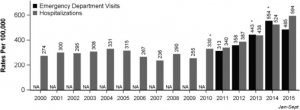
Dr. Wang and his team conducted a retrospective review of data collected from the Colorado Hospital Association (CHA) Discharge Databank and National Poison Data System (NPDS) data repository. The primary objective was to compare rates of hospitalizations and ED visits with marijuana-related billing codes and RPC calls with mention of marijuana. The secondary objective was to compare the primary diagnosis categories of hospitalizations and ED visits with marijuana-related billing codes to those without.

From 2000 to 2015, hospitalization rates with marijuana-related billing codes increased from 274 to 593 per 100,000. Interestingly, the prevalence of mental illness among ED visits with marijuana-related codes was 5x higher than the prevalence without – potentially due to the subsection of patients often receiving more detailed assessment around substance abuse habits, or having a greater number of visits to the ED. RPC calls remained constant from 2000 to 2009, but upon the legalization of medical marijuana in 2010 the number increased from 42 to 93. After recreational legalization in 2014, RPC calls increased from 123 to 221.

Figure 1: Rates of hospitalizations and ED visits with marijuana-related billing codes in the first three diagnosis codes in CO

Figure 2: Annual RPC human exposure calls related to marijuana in CO, by age group

Overall, legalization of both medical and recreational marijuana in Colorado is associated with an increase in hospitalizations, ED visits, and RPC calls. Since the study was conducted in a state with legalized recreational marijuana, the findings are likely generalizable to states with similar laws. However, as expected in such complex area, limitations do exist. Researchers relied solely on ICD codes and drug screens without conducting full chart reviews. Marijuana use cannot be viewed as the direct cause of most ED visits, hospitalizations, or RPC calls, as the effects are often short-lasting and not clinically relevant. The data provided does not establish causality between hospitalizations or ED visits secondary to marijuana exposure, and future research in this area should emphasize the importance of not overestimating the healthcare burden related to marijuana use. While researchers did well to establish an association between legalization of marijuana and increased healthcare utilization, there is still much work to be done in this area.

Key Terms

  • Marijuana: colloquial term for the flowering plant intended for consumption. Used in discussion of Wang et al. to align with manuscript terminology.
  • Cannabis: also known as marijuana, is the science-based term of flowering plant intended for consumption.
  • Cannabinoid: a single class of diverse chemical compounds that act on cannabinoid receptors in the brain. The most notable cannabinoid is tetrahydrocannabinol (THC), the primary psychoactive compound in cannabis.
  • Dronabinol: the international nonproprietary name (INN) for a pure isomer of THC, often used to treat anorexia in patients with HIV/AIDS as well as for refractory nausea and vomiting in people undergoing chemotherapy.
  • Nabilone: a synthetic cannabinoid that mimics THC and is often used as an antiemetic and adjunct analgesic for neuropathic pain.

[i] CBHSQ (Center for Behavioral Health Statistics and Quality). 2016. Behavioral health trends in the United States: Results from the 2014 National Survey on Drug Use and Health (HHS Publication No. SMA 15-4927, NSDUH Series H-50). 

[ii] NCSL (National Conference of State Legislatures). 2016. State medical marijuana laws.

[iii] Pines JM. – Cannabis in the Emergency Department: policy vs. evidence with Dr. Esther Choo. Urgent Matters Podcast. 2017

[iv] National Academies of Sciences, Engineering, and Medicine. 2017. The Health Effects of Cannabis and Cannabinoids: The Current State of Evidence and Recommendations for Research. Washington, DC: The National Academies Press. doi:https://doi.org/10.17226/24625.

[v] Wang GS, Hall K, Vigil D, Banerji S, Monte A, VanDyke M. Marijuana and acute health care contacts in Colorado. Prev Med. 2017 Mar 30. pii: S0091-7435(17)30120-2. doi: 10.1016/j.ypmed.2017.03.022.


Ameer Khalek is a MPH student at the GWU Milken Institute School of Public Health

Greg Jasani

August 2, 2017

Febrile neutropenia is a well known side effect of chemotherapy and patients with it often present to emergency departments.  Standard of care has been to admit these patients for aggressive IV antibiotics. However, recent research has suggested that a certain subset of these patients can be considered low risk and managed as outpatients[1],[2].  Two risk stratification tools, the Multinational Association for Supportive Care in Cancer (MASCC) and Clinical Index of Stable Febrile Neutropenia (CISNE) scores, have been proposed to identify these low risk patients[3].  While these scores have been studied and validated in the inpatient setting, there has been no research about whether these risk stratification tools are valid in the emergency department.  A recent article in Annals of Emergency Medicine by Coyne et al examined whether these scores could be used to accurately identify low-risk febrile neutropenic patients in the emergency department.

The study was a retrospective cohort study conducted in the emergency departments of two academic hospitals.  It examined the medical records of all patients who presented with febrile neutropenia to the two emergency departments between June 2012 and January 2015.  Inclusion criteria for the study were a fever of greater than 100.4F and an absolute neutrophil count of less than 1,000.  Only patients with febrile neutropenia believed to be secondary to chemotherapy were included.  In total, the authors reviewed 230 charts in their final analysis.  The primary outcomes of the study were inpatient length of stay, level of care, clinical deterioration, clinical deterioration, positive blood cultures, and death.  For the purposes of the study, the authors considered patients who did not experience any negative outcomes during their admission to be truly low risk.

Source: Coyne et al.  Application of the MASCC and CISNE risk-stratification scores to identify low-risk febrile febrile neutropenic patients in the emergency department. Ann Emerg Med.  2017; 69(6): 755-764

For every patient, the authors calculated both the CISNE and MASCC scores to determine which was more accurate in identifying low-risk patients (See the Figure for details on how to calculate these scores).  Ultimately, they found that the CISNE score was more accurate in identifying febrile neutropenic patients in the ED did not have an adverse event while hospitalized.  The score was 100% specific in identifying all negative outcomes except for positive blood cultures for which the specificity was 97.4%.  The positive predictive value of the CISNE score in the identification of low risk patients was found to be 98.1%.  The MASCC score was less accurate at identifying low risk patients.  The specificity for identifying low risk patients was only 54.2% and the positive predictive value was 84%.

This study provides evidence that the CISNE score can be used to accurately identify low-risk febrile neutropenic patients in the emergency department.  The CISNE score was also found to be superior to the MASCC score.  Although this study was a retrospective chart review, it provides evidence that the CISNE score may be helpful in determining which patients are at low risk of having adverse outcomes associated with their condition.  There is growing evidence to suggest that these patients can be managed successfully as outpatients[4],[5].  Managing them as outpatients would not only save hospital resources but prevent exposing these patients to potentially harmful hospital acquired infections.  While this paper is not enough justification to discharge these patients directly from the emergency department it does show that further research into this area is warranted and provides a potentially helpful risk stratification tool for emergency medicine physicians to help guide clinical decision making.

[1] Cooksley, T., Holland, M., and Klastersky, J. Ambulatory outpatient management of patients with   low risk febrile neutropaenia. Acute Med. 2015; 14: 178–181

[2] Gea-Banacloche, J. Evidence-based approach to treatment of febrile neutropenia in hematologic malignancies. Hematol Am Soc Hematol Educ Program. 2013; 2013: 414–422

[3] Coyne C, Le V, Brennan J, Castillo E, Shatsky R, Ferran K, et al.  Application of the MASCC and CISNE risk-stratification scores to identify low-risk febrile febrile neutropenic patients in the emergency department. Ann Emerg Med.  2017; 69(6): 755-764

[4] Flowers, C.R. and Karten, C. Communicating safe outpatient management of fever and neutropenia. J Oncol Pract. 2013; 9: 207–210

[5] Pherwani, N., Ghayad, J.M., Holle, L.M. et al. Outpatient management of febrile neutropenia associated with cancer chemotherapy: risk stratification and treatment review. Am J Health Syst Pharm. 2015; 72: 619–631


Greg Jasani is a fourth year medical student at the GW School of Medicine & Health Sciences

1

Austin Wu

July 17, 2017

A patient presents to the emergency department presenting with precordial chest discomfort, pain radiating to the jaw, dyspnea, and diaphoresis. These are some of the typical symptoms highly indicative of acute coronary syndrome (ACS) [i]. However, not all patients have “typical” symptoms. Prior studies have shown that certain demographic groups, specifically patients of older age, black or Asian race, and female gender are less likely to present with these typical symptoms despite later having the diagnosis of ACS [ii].  In addition, other factors have been associated with atypical presentations including older age, and the presence of comorbid conditions such as diabetes.

While there is a large literature on atypical presentations of ACS, the combination of demographic factors – specifically combinations of race and gender – has not been closely explored until now. A recent study by researchers Drs. Ahmed Allabban, Judd Hollander and Jesse Pines at the George Washington University School of Medicine and Thomas Jefferson University published in the Emergency Medicine Journal focused on the combination of race and gender, and how the intersection of these two factors correlate with presentation of 30-day ACS or other serious cardiopulmonary diagnoses [iii]. The four subgroups were analyzed: black males, white males, black females, and white females.

The study was a secondary analysis of data collected from a prospective, observational cohort study of ED patients presenting with chest pain, with timeframe spanning from 1999 to 2008. The entire dataset was more than 4000 patients, and the study was conducted at an inner city academic hospital. Inclusion criteria included 30 years of age or older, presentation of chest pain where an ECG was ordered, and provision of informed consent. Patients who had chest trauma within the past week, a measured temperature of 101°F, used home oxygen, had metastatic cancer or had symptoms of palpitation alone without chest pain were excluded from the study.

The study found that symptoms associated with a higher risk of ACS in white males were left arm radiation, chest pressure and tightness, and substernal chest pain. These are typical symptoms of ACS.  For black males, the symptom associated with a higher risk of ACS was diaphoresis. For black females, symptoms indicating higher risk of ACS included diaphoresis, palpitations, and left arm radiation, while symptoms indicating lower risk for ACS included pleuritic chest pain and left anterior chest pain. No symptoms were predictive of ACS for white females. For serious cardiopulmonary diagnoses, there were largely similar findings. Results are displayed in the figure below.

The data demonstrates that dividing patient populations by a combination of race and gender produces variably predictive symptoms of both ACS and other serious cardiopulmonary diagnoses. Thus, relying on a particular set of typical symptoms regardless of demographic representation may not be an optimal form of practice.

This study had several limitations, including being a single center study, using convenience sampling, and having variations in sample size for each subgroup. Further, while this study highlights differences in symptoms among race and gender subgroups, the question of what causes these differences remains an area to be explored. Preliminary explanations include hormonal, physiological, and sociocultural differences among each of these subgroups, though these have not been substantiated. The main takeaway is that physicians in the ED should keep these symptomatic differences between race and gender in mind when assessing a patient for chest pain.

[i] Ayman El-Menyar et al., “Atypical Presentation of Acute Coronary Syndrome: A Significant Independent Predictor of in-Hospital Mortality,” Journal of Cardiology 57, no. 2 (March 2011): 165–71, doi:10.1016/j.jjcc.2010.11.008.

[ii] H. Lee et al., “Typical and Atypical Symptoms of Myocardial Infarction among African-Americans, Whites, and Koreans,” Critical Care Nursing Clinics of North America 13, no. 4 (December 2001): 531–39.

[iii] Ahmed Allabban, Judd E. Hollander, and Jesse M. Pines, “Gender, Race and the Presentation of Acute Coronary Syndrome and Serious Cardiopulmonary Diagnoses in ED Patients with Chest Pain,” Emerg Med J, June 16, 2017, emermed-2016-206104, doi:10.1136/emermed-2016-206104.


Austin Wu is a medical student at the GW School of Medicine & Health Sciences

Caitlin Carter

July 13, 2017

Antimicrobial overuse is the most significant preventable cause of drug resistance in both hospital and community settings[i].

Antibiotic overuse occurs among practitioners in nearly 2/3 of cases.

Overuse contributes to the growth of antibiotic resistant pathogens and the spread of antibiotic resistant infections that lead to increased morbidity, mortality, and health care costs.

Antimicrobial stewardship has emerged as a solution to combat this dangerous and preventable threat to patient safety. Antimicrobial stewardship is a collection of policies, guidelines, surveillance, data transparency, education, and evaluation to optimize antibiotic-prescribing practices. These strategies can lead to better patient outcomes, a decrease in adverse events and infections, and an optimized use of resources.

Last year the Infectious Diseases Society of America and the Society for Healthcare Epidemiology of America released a set of evidenced-based guidelines on antimicrobial stewardship in clinical settings[ii]. These guidelines serve to improve and measure the appropriate use of antibiotics across dosing, duration of care, and route of administration. While these guidelines were developed by a panel of clinicians and investigators across various medical disciplines.

Emergency physicians have a unique vantage point to affect antimicrobial stewardship in inpatient and outpatient settings.

The initial choice of antibiotic in the Emergency Department (ED) has important downstream implications. ED clinicians routinely prescribe antimicrobials for patients with skin and soft-tissue infections, urinary tract infections, bloodstream infections, as well as upper and lower respiratory infections. Broad-spectrum antibiotics are sometimes overused in EDs and other ambulatory settings, and in observational studies have shown significant overprescribing for acute bronchitis and other conditions.

Antimicrobial stewardship in ED settings is particularly challenging due to the high rates of ED crowding, the rapid rate of patient turnover, the need for quick decisions without consultation, the shift-based scheduling format of providers, and higher staff turnover rates than in other clinical settings. In addition, there are provider-centered factors that impact stewardship. This includes a perceived lack of efficacy, concerns about resource availability and reimbursement, as well as perceived hindrance to operational efficiency. Providers are also concerned about medical liability and patient satisfaction, which has shown to be an important facet of antibiotic prescription in the ED.

Challenges to antimicrobial stewardship in ED settings are unique, numerous, and require strategies that consider the ED as well as other key stakeholders. ED specific guidelines are still needed that consider clinical decision support systems, post prescription review, rapid diagnostics, duration of therapy, dosage, and ED antibiogram development.

To learn more about antimicrobial stewardship and implications for emergency medicine please see Dr. Larissa May’s podcast and webinar available only through Urgent Matters. For an overview of antibiotic use in pneumonia, please see Dr. Jesse Pines’ webinar on the burden of community-acquired pneumonia and current issues in emergency care.

These programs were funded through an educational grant from Cempra, Inc. (CEMP) who had no role in the content.

[i] May, L., Cosgrove, S., et al. “A Call to Action for Antimicrobial Stewardship in the Emergency Department: Approaches and Strategies.” Annals of Emergency Medicine (2013).

[ii] Barlam, T.F., Cosgrove, S.E., et al. “Implementing an Antibiotic Stewardship Program: Guidelines by the Infectious Diseases Society of America and the Society for Healthcare Epidemiology of America.” Clinical Infectious Diseases (2016).


Caitlin Carter, MPH is a research associate and manager for the Urgent Matters program