Objectives:
- Identify microscopic structures of the thyroid, parathyroid, and adrenal glands.
- Identify specific cells/structures of each gland:
- Thyroid- follicular epithelial cells, parafollicular cells (C cells)
- Parathyroid- chief cells and oxyphil cells
- Adrenal- cortex, medulla, zona glomerulosa, zona fasciculata, zona reticularis, chromaffin cells
Overview:
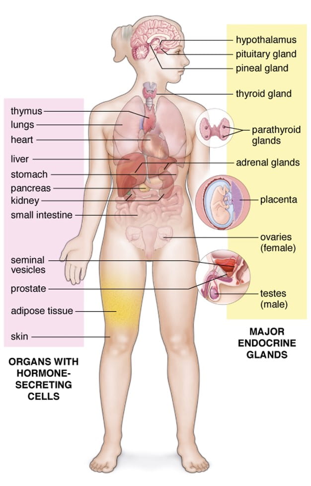
The general purpose of the endocrine system is to produce hormones that circulate throughout the body via connective tissues (CT) and the vascular system to regulate function. Endocrine glands are generally structured as aggregates of epithelioid cells embedded in connective tissue. There is no duct system, so hormone products are secreted through the CT into capillaries. The endocrine system involves major endocrine glands and hormone-secreting cells found in other tissues. In general, endocrine glands are associated closely associated with fenestrated capillaries, ensuring rapid entry of hormones into the systemic circulation. The image below demonstrates how the endocrine system is spread throughout the body. This page will cover the thyroid gland, parathyroid glands, and adrenal glands. Other organs are covered in different pages of this website.
Thyroid Gland:
The thyroid gland is located in the neck region, anterior to the larynx and trachea. This bilobed organ forms the shape of a butterfly with two lateral wings/lobes connected by an isthmus (thin band of tissue). In some cases, there is a pyramidal lobe which extends superiorly from the isthmus. The general microscopic structures consists of spherical, cyst-like follicles filled with colloid (gel-like mass) embedded in CT. The follicles, functional units of the gland, are lined by simple cuboidal-low columnar epithelium resting on a typical basal lamina.
Cells-
- Follicular cells (principal cells)- epithelial cells that vary from cuboidal to columnar in height with slightly basophilic cytoplasm, spherical nucleus and produce T4 and T3 hormones.
- Parafollicular cells (C cells)- located in the follicular basal lamina or in adjacent CT. They are pale-staining and secrete calcitonin. Can be difficult to identify in typical light microscopy.
Parathyroid Glands:
Parathyroid glands are small, ovoid glands found in 2 pairs located in the CT of the posterior surface of the lateral lobes of the thyroid gland. One pair is superior and the other pair is inferior. Some people have more or less than a total of 4 glands. The gland is poorly divided by CT septa into lobules which contain densely packed cords of cells.
Cells-
- Principal (chief) cells– small polygonal epithelial cells with a centrally located nucleus and slightly acidophilic cytoplasm. They Regulate synthesis, storage and secretion of parathyroid hormone (PTH). They are found in larger in number compared to oxyphil cells.
- Oxyphil cells– larger, rounded cells with an acidophilic cytoplasm. Found in smaller numbers than principal cells. They have no known secretory role.
Adrenal Glands:
Adrenal glands are paired glands found in the retroperitoneal space of the abdominal cavity located just superior to the kidneys but do not participate directly in kidney function. The right gland is triangular while the left gland is semilunar. They secrete steroid hormones and catecholamines. Each gland is surrounded by a CT capsule and contains a cortex, a steroid secreting region, and a medulla, the catecholamine-secreting region.
Zones of the cortex:
- Zona glomerulosa– narrow, outer zone, that secretes aldosterone and mineralocorticoid. Cells in this zone are small, columnar-pyramidal shape with spherical nuclei and foamy cytoplasm.
- Zona fasciculata– thick, middle zone, that secrete glucocorticoids. Cells are large and polyhedral arranged into straight cords with foamy cytoplasm, 1-2 cells thick separated by sinusoidal capillaries.
- Zona reticularis– thin, inner zone, secretes gonadocorticoids (adrenal androgens). Cells are Small, light and dark-stained cells with deeply-stained nuclei arranged in cords separated by fenestrated capillaries.
Cells and Structures of the Medulla:
- Chromaffin cells– large, pale-staining epithelioid cells organized in cords and innervated by presynaptic sympathetic neurons. These cells are basically, modified nerve cells lacking axonal processes. They secrete catecholamines.
- Ganglion cells– large cells with a round nucleus and prominent nucleolus.
- Blood and Lymphatic vessels
Endocrine Gland Slides

This specimen of the thyroid lacks the dense CT capsule but you can see the individual follicles filled with colloid. Study the epithelium and identify the primary follicular cells and look for parafollicular cells.

Use this specimen slide to study the microscopic features of the parathyroid gland. In the adult, this gland develops adipocytes which can constitute up to 70% of the glandular mass.

The adrenal is surrounded by a thin CT capsule and consists of a cortex and medulla. The cortex can be further subdivided into zones: zona glomerulosa, zona fasciculata, and zona reticularis. In the zona reticularis, look for lipofuscin pigment in some of the cells. In the medulla, you will find chromaffin cells filled with granules and ganglion cells which have a larger nucleus with prominent nucleolus. You will also see extensive vasculature.
